


Case presentation:
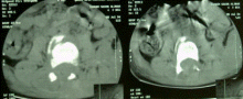
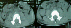
A 4 years old girl after suffering RTA 8-7-2004, got disruption of the vertebral column at L3 due massive traction with the seat belt in place. On inspection, the flank was full of subcutaneous hematoma and she was paraplegic at the level of L1. The patient was conscious and plain X-rays and CT-scan abdomen and vertebral column showed the flail vertebral column at the level of L2-3.
The patient was operated 8-7-2004: Skeletonization of L2 & L3 was performed. There was a big gap between the two spinous proccessi. The lower extruding backward with all the ligamentous structures ruptured. The facets of L2 were seen lying more superficial than the lamina of L3. The dura was missing at the level of the defect. No CSF was coming out. and the visible roots were intact. Intentionally, no attempt was performed to explore the neural structures. Open reduction of the locked facets was done, after what the bony alignment regained more or less, acceptable appearance, but lacking stability, for what, the smallest available Hartchell rectangle was used. It was 4 cm length and aided with sublaminar wires. Considering that the girl is only 4 years age and the bone is relatively soft, the sublaminar wiring was performed, using the smallest diameter wires bundled in 4 rows, then turned, to achieve a wire of 8 wires, were used to fix the construct to the L2 and L3 laminae. This trick was used by me, for several years in osteoporotic patients, where their bone cannot tolerate the use of strong big diameter wires. The gap between the two spinous proccessi diminished and it was covered by muscle, to prevent CSF leak and tight closure of the wound performed.
![]()