NEW SURGICAL APPROACH TO THE
CLIVO-PONTO-CEREBELLAR ANGLE REGION
Dr. M. A. Elias M. D., Ph.D.
Head of Neurosurgical Department
AI-Bashir Hospital. MOH.
Objectives: A new method was introduced during surgical approaches to the clival and the ponto-cerebellar region, by using the high speed economic staged drilling of the temporal bone, so that the subtemporal trans superior petrosal sinus I retro-antesigmoid transtentorial approach to the clivus can be gained without massive destruction to the fine important anatomic structures of the inner ear.
Patients and Method: Starting from January 1995 we started to change our surgical tactics, concerning patients with giant tumours of the clivus and the ponto-cerebellar angle. Using the standard approaches proposed by U. Fisch and the others, we noticed the grave complications arising from the dissection of the superficial petrosal nerve and the unavoidable hearing loss and the facial paralysis despite anatomical preservation related with such procedures. We found the necessity to resolve these problems and 18 patients were operated using the new technical standards introduced in our department.
Results : 18 patients were operated during 1995-1997 using the approaches to the clivus and ponto-cerebellar angle. 10 females and 8 males within the age of 8 -56 years with mean age 38± 6 years. 8 of them suffered of giant acoustic neurinoma and 6-meningiomas, 2 epidermoids I - exophytic astrocytoma and one of retrosellar clival craniopharyngioma.
Postoperative mortality was I 1. I % which was not related directly to the type of surgical attack. 4 patients with hearing loss with epidermoid and clival meningiomas before operation, regained hearing immediately after operation. Exposure keratitis, which was previously the common concomitant manifestation after the proposed earlier operations disappeared in our series of patients. It was only noted at the first operation performed with relatively major dissection anterior to the internal meatus acousticus. Facial paresis took place in 5 patients, where dissection of the facial nerve at the internal auditory canal was performed. This paresis was minimal in the first postoperative day, to increase in intensity in the second postoperative day.
Conclusion: It is not necessary to perform radical petrosectomy in each case with tumours invading the clivus and the ponto-cerebellar angle. Even in the case that the tumour was extending to the inner ear structures, it is not necessary to drill out all the minute delicate structures of the inner ear. Surgical planning must be directed at minimal resection to obtain the visual access to the tumour boundaries and not to work in other angles, most of which are needless. The mastoid cavity was not attacked most of the time. The inner ear cavity in I 0 cases was left intact even in the case that the vestibular schwannoma was destroying the meatus acousticus iternus. The bony fallopian canal was left undissected. Dissection was done in the case of direct invasion of the nerve at that site. Partial medial vestibulectomy was performed if needed to gain access to the internal auditory canal and foramen magnum.
At AI-Bashir Hospital during the last 10 years more than 750 operations were done for the various histological types of brain tumours with various localization. Many attempts were made to refine the technical standards in neurooncology. We started to use the techniques described by Ugo Fisch since 1988, Presence of hearing was an obstacle to perform such a procedure. Then several techniques emerged describing some certain routes, repeating what House W.H. in the past proposed with some modifications. We used all these standards, but after drilling of the temporal bone all the patients were forced to pay certain complications, related to these types of approaches. Most of them developed exposure keratitis due to trauma to the superficial petrosal nerve, paralysis and deep paresis of the facial nerve due to its bony dissection and transposition and the eminent loss of hearing recovery in disputable cases.
During the last 3 years we started to approach the pontocerebellar angle and the clivus with planned staged drilling, starting with the creation of the wide osteoplastic craniotomy in the temporo-occipital region abutting the horizontal plane of the middle fossa. By using the high-speed drill we bevelled the surface of the middle fossa starting to expose the sigmoid sinus as a landmark, then we proceeded to expose the superior petrosal sinus to an extent of 20-25 mm, which was usually sufficient to clip the sinus. Resection of the lateral edge of the tentorium was graded with the necessity of the tumour relationships with the tentorial hiatus. If the tumour was transgessering the tentorium, then complete tentoriotomy was done. In other cases partial tentoriotomy was performed to gain visual control to the lesion. Our material is relatively small in number, but we did not have to ligate the sigmoid sinus, even in the presence of caudal extension of some tumours down to the level of the second cervical spine. Drilling must be wide -surfaced with the aim to remove the sharp angles, obstructing the line of vision between the surgeons eyes and the boundaries of the tumour. In the presence of intracanalicular extension of the tumour a transvestibular approach was extended. To gain adequate approach to that part. 2 deaths occurred in our series with clival meningiomas of giant dimensions. The death happened 3 and 6 months after operation due to problems related either to the respiratory tracts and centre, the caudal group of nerves, despite their anatomical preservation, suffered more functional decline postoperatively. The tumours in all cases were radically removed.
Case presentation:
Case 1.: A patient, an engineer, 35 year age came to us complaining of inability to walk steadily and regurgitation during swallowing for several months, increasing gradually with headache and blurring vision. MRl showed a giant clival meningioma distorting the brainstem, as it is the cover of the tumour. A right subtemporal trans-superior petrosal sinus, transtentorial, retro-antesigmoid approach was done 15.5.1996 as described and the tumour was radically removed. Postoperatively the patient was sent to ICU, and he regained his consciousness the 2-d postoperative day. The patient progressed shocked lung the next few days. Tracheostomy was done the 5-th postoperative day. During the next week after failed CVP line insertion, he developed Rt sided heamopneumothorax, which was seemingly minimal in routine X-ray studies. Chest CT-scan was requested 10 days later, showing pyopneumothorax Rt lung, for what chest tube was inserted. The patients breathing drive improved, after what it was possible to disconnect him off ventilator. The chest tube was removed 2 days ago. Control CT-scan showed still persisting cavity without pus collection, but with thick capsule.
Considering that, our hospital had no thoracic surgeon at that time. The patient was transferred to the Islamic Hospital, at the request of his relatives. Decortication of the pleural adhesions was done, after what his respiratory functions improved. Three days after discharge from there, he was readmitted to our hospital, with clinical picture of tracheal stenosis at the tracheostomy site. The wound was almost closed. The tracheostomy was reinserted and his difficulty in breathing considerably disappeared. Three days after discharge he was readmitted to our hospital, with clinical picture of tracheal stenosis at the tracheostomy site. The wound was completely closed. The tracheostomy was reinserted and his difficulty in breathing considerably disappeared. The patient, despite the preservation of the caudal group of cranial nerves, still suffering of swallowing difficulties, which usually needs about 5-6 months to regain its functional recovery. There was also exposure keratitis Rt eye. The patient was then in TPN, receiving maalox 10 ml every 3 h. Tienam I gm x8h, Amikin 500 mg x8h, chloramphenicol I gm x8h, Tagamet 200 mg x8h. Attempt for NGT feeding 3 days ago triggered the stress ulcer, for what, it was stopped another time. The patient under continues treatment could regain his walking abilities and he was stable, except for the respiratory functions, which were deteriorating when Tienam was absent in the hospital and several CXS done periodically showed response only to Tienam. The patient within three days deteriorated and died with signs of tracheo-pulmonary obstruction 5. September. 1996. This patient developed exposure keratitis of the right eye due to massive drilling anterior to the internal auditory meatus and possibly traumatizing during that, the superficial petrosal nerve.
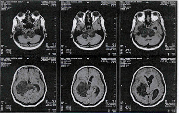
Case 14 : The patient was a young girl 29 years old came from the Palestinian self-rule area to our department complaining of severe headache for more than 8 months with tinnitus right ear for 3 months, inability to walk without aid for 2 months. The patient was fully conscious with depressed mood. Her higher mental functions were preserved Neurologically, the patient had blurring vision with decreased hearing both sides. There was considerable facial paresis left side with paresis of the external rectal muscle left eye. Romberg testing was impossible, because the patient was unable to stand in upright standing position. There was horizontal nystagmus when looking to both sides.

Case 14 MRI showing the dimensions of the epidermoid
CT-scan and MRI done the day before admission, showing hypointense mass in TW I sequences ad hyperintense in TW2. The mass was suggested to be a dermoid.
Considering the morphological picture of the mass and the great dimensions, it was decided to be attacked from the right subtemporal route. I I. December. 1996: Osteoplastic craniotomy in the right fronto-temporal region was done. The flap was extended far inferiorly abutting the zygomatic arch. The flap was rotated to the direction of the ear. Economic drilling of the superior parts of the temporal bone abutting the trajectory of the superior petrosal sinus and the sigmoid sinus. The dura was very weakly transmitting the cardiopulmonary pulsation. The dura was opened parallel to the inferior edge of the bony defect. Clipping of the superior petrosal sinus and partial tentoriotomy with antesigmoid approach was done. The thinned layer of the right temporal lobe was resected. It was about 5 mm in thickness, below which the tumour started to appear. It has the pearl-la-mutter appearance with cholesteatoma substance easily succable. Through a 2X2 cm visual access it was possible to remove the entire mass that was located supratentorialy. The tentorial superior surface was not attached firmly with the mass. The isocenter of the mass was debulked, after what the basilar artery and bifurcation started to appear in the field. The mass was engulfing and in other places encasing the basilar, PCAa, the CCAa, both Pc0Aa, AICAa, PICAa and the vertebral arteries. The tumour was not infiltrating the pons or the medulla, but in the interpeduncular cistern its capsule was adherent to the cerebral peduncles. Meticulous peace-meal resection was done in that area, preserving during that the perforating arteries, which were stretched by the pockets of the arachnoid containing the dermoid material. The material was dissected off the mammilary bodies and both oculomotor nerves, which proved to be elongated and thinned by the pathologic process. They were completely engulfed by the white avascular substance. The vascular structures were hanging freely away from the brain stem, after removal of the entire mass between them and the brain stem. The mass was growing far posteriorly along the supratentorial space, reaching the right occipital pole. This part was resected preserving during that the draining veins. That part that was reaching the quadrigeminal cistern to the contralateral side was followed and resected. It was with considerable attachment with the deep cerebral vein ipslateral side. Subtentorially the tumour was followed parallel to the clivus, during removal of which the vein of Dendy right side was preserved. The clivus came to visual control, after what it was possible to identify both abducens nerves and the contralateral elongated trigeminal nerve. The entire foramen magnum structures were under visual control. It was possible to see the spinal cord running down up to C3 level.
Postoperative course was smooth: the patient regained her consciousness immediately after operation, extubated with no apparent sensori-motor deficit. The abducens paresis of the left eye deepened. Considering her preoperative swallowing deficits with regurgitation, it was decided to keep her in NGT for 2 days, and then she started normal feeding. Discharged to be in Epanutin 100 mg for 18 months.
Case No 16. The patient was a young female 25 years old, married, complaining of severe headache and loss of hearing with tinnitus for the last 2 years. Diplopea with trigeminal pain at the distribution of the right VI for the same period of time. The patient was not known to have diabetes mellitus or arterial hypertension. MRI was done 6.Mar. 1997 at the Islamic hospital showing the schwannoma extending infratentorially with dimensions 4X4X4 cm. It is iso-intense in T1W images and becoming hyper-intense after contrast with gadolinium. MRA was done showing a small berry aneurysm at the basilar tip bifurcation.
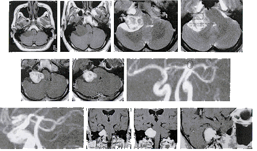
Audiometry was done indicating severe to profound sensory neural deafness in the right ear. The patient had hypalgesia of the right VI distribution. Intraocular ophthalmoplegia with skew deviation. The right eye looked to the right when asking the patient to look forward. The convergence and accommodation were disturbed. Romberg test was: the patient had tendency to sway to the right. There were no data supporting any signs of previous rupture of the berry aneurysm. The right facial nerve was intact functionally. Considering the giant dimension of the tumour and the lost hearing right car and the presence of berry aneurysm at the tip of the basilar artery bifurcation, it was advisable to attack the tumour through the right trans-petrosal sinus ante-sigmoid approach.
Operation was performed 23.3.1997. Through right ante-sigmoid translabirinthine transmeatal approach with clipping and separation of the superior petrosal sinus and partial resection of the tentorium the tumour was identified in its intracanalicular portion. The tumour had dumbbell-shaped appearance with the waist is constricted by a dural ring and adherent to it. The tumour was highly vascular firm in consistency. It had a well-defined cleavage. The intracanalicular part was resected and the dural attachments were followed and resected. A dural defect about 2OX30 mm was created mainly in the antesigmoid posterior fossa portion of the dura, which consisted the medial wall of the pyramid behind the meatus acousticus iternus. Intracapsular piece-meal resection of the intracranial part of the tumour was done.
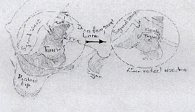
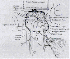
Schematic drawing showing the relationship of the acoustic neurinoma to the surrounding anatomic structures.
After decreasing the tumour in size, it was possible to dissect its capsule off the tentorium, the vein of Dendy and the facial nerve, which was pushed antero-superiorly and the trigeminal nerve, which was pushed infero-anteriorly and some branches of the caudal group of nerves, including the accessory nerve. AICA was dissected sharply and preserved. After that the remnant of the tumour attached to the brain stem was followed with sharp dissection and totally removed including its origin in the right acoustic nerve. Physiologic parameters during manipulation in the brain stem were acceptable. Considering the giant dimensions of the tumour and possible evolution of heavy complications in the postoperative course it was decided not to touch the berry aneurysm. The dural defect and the extradural cavity created after bony dissection was filled by a muscle obtained from the quadriceps muscle right leg. Routine closure. Postoperative immediate recovery was uneventful. The facial nerve was paretic, but functioning, which increased in severity in the second postoperative day.
Discussion:
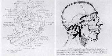
Various surgical approaches to the posterior cranial fossa through the temporal bone are available. Entry through the temporal bone imposes special problems if the internal carotid artery, sigmoid sinus, cranial nerves VII and VIII, and the specialized structures for hearing and balance are to be preserved. The transtemporal approaches to the posterior fossa are classified into two groups: (1) the basic individual approaches and (2) combined approaches.

Ojemann R.G. stated that, both the suboccipital and translabirinthine approaches have been used to remove tumours of all size. When hearing is to be preserved, the suboccipital approach is preferred. Some surgeons use the middle fossa approach for small tumours in the internal auditory canal, particularly at the lateral end when an attempt is being made to preserve hearing. The surgeon must have a thorough understanding of the anatomy of the cerebellopontine angle and petrous bone and the relationship of anatomic structures to the tumour. In transmeatal suboccipital approach a vertical incision is centred I to 2 cm medial to the mastoid process. A graft of pericranial tissue about 4 cm in diameter is taken from the occipital region by extending the superior aspect of the incision as needed. This graft is used in closing the cerebellar convexity dura at the end of the operation. The suboccipital muscles and fascia are incised in line with the incision and are carefully separated from their attachments to the bone. The bone over the lateral two thirds of the cerebellar hemisphere is exposed. Visualizing the midline bone or rim of the foramen magnum is usually not necessary. A borehole is placed, and the dura is carefully separated from the overlying bone, and a bone flap is cut. The mastoid air cells are usually entered and are occluded with bone wax. In some patients with large tumours, a small portion of the lateral cerebellar hemisphere is removed to facilitate exposure. After revision the eighth nerve complex, the dura over the region of the internal auditory canal is removed and bone is removed by drilling. When hearing preservation is a consideration, bone removal extends for a distance of no more than 10 mm laterally. A more lateral exposure risks entering the labyrinth, which is usually associated with loss of hearing. However, in some patients immediate occlusion of the open labyrinth with bone wax allow hearing to be retained.
The petrosal vein, which usually comes off the cerebellum or middle cerebellar peduncle to the petrosal sinus just above the tumour, is coagulated and divided. After the completion of tumour removal the area of bone removal over the internal auditory meatus is carefully waxed, and adipose graft taken from a superficial incision on the lower abdomen is packed there.

The translabirinthine approach has been introduced by House W. in 1960. As King T.T. et al counted its contraindications, mentioned the excellent exposure of the lateral end of the meatus and the allowance to the facial nerve to be identified as inters the fallopian canal. An absolute advantage is that the facial nerve can be exposed throughout its petrous course, a useful feature if the tumour arises from that nerve and extends along into the geniculate ganglion and beyond. Mobilization of the facial nerve out of its bony canal is useful if the nerve is damaged during the excision of the tumour and needs reconstruction. The main advantage of the translabirinthine method is the direct approach it offers to the cerebellopontine angle. The semicircular canals and the vestibule lie directly between the mastoid air cell system and the internal auditory meatus and must be removed together with the endolymphatic duct to expose that canal. The facial nerve can always be found in the lateral end of the meatus, where it inters the fallopian canal and is found immediately anterior to the superior vestibular nerve, separated from it by a narrow vertical osseous septum (Bill's bar). The bony dissection begins in McEwan's triangle, posterior to the spine of Henle. The lateral mastoid cortex is removed, and the immediate landmarks exposed are the middle fossa plate superiorly and the sigmoid sinus plate posteriorly. Particular care is taken to extend bony removal superiorly over the adjacent lateral surface of the temporal bone and posteriorly behind the sigmoid sinus. In these two areas, extra room is therefore made available, and this room is critically important because by increasing the superficial limits of the exposure, the arc through which the surgeon may work in depth of the cerebellopontine angle is greatly increased. Thus if the temporal lobe dura is elevated, the inferior pole of the tumour can be visualized, and If the sigmoid sinus is retracted backward and collapsed, the posterior face of the petrous bone medial to the meatus and up to the trigeminal nerve and tentorial hiatus can be approached. If additional room is needed, bone may be removed superiorly over the temporal lobe, with a punch forceps. The bony dissection continues medially, and the Koerner septum is removed before important landmark of the lateral semicircular canal is visualized on the medial wall of the epitympanic recess. The lateral canal, characterized by its ivory-like appearance and lack of pneumatization, is the key to the position of the facial nerve, which lies inferiorly and deep to the most lateral part of its arch. The mastoid ant= is widely opened and the whole mastoid cavity is expanded. A shell of bone is left over the middle fossa dura, the sigmoid sinus, and the sino-dural angle at this stage because venous bleeding from these sites may interfere with the ease of deeper dissection. Complete unroofing of the sinu-dural angle is very important in the final exposure. Attention is now paid to the epitympanum, which is reached by thinning of the posterosuperior wall of the external auditory meatus until the body of the incus and the head of the malleus are visible. The incus may now be excised by rotation of the short process superiorly. Disarticulation from the malleus and the stapes can be achieved without subluxation of the latter from the oval window. The facial nerve is now clearly visible and may be followed posteriorly to the second genu and then, by careful bone removal, skeletonized as it descends through its mastoid segment to the stylomastoid foramen. The digastric groove is then uncovered, thereby clearly locating the stylomastoid foramen and clearing bone between the sigmoid sinus and descending facial nerve. Attention is now turned to the creation of a posterior tympanotomy. Bone is removed between the anterolateral face of the facial nerve medial to the fibrous annulus of the external auditory meatus and the chorda tympani. Bone removal continues, although it may be constrained by the position of the cauda tympani, until the round window niche is visible. Such a dissection gives a view adequate to check the footplate of the stapes and the oval window and to obliterate the middle ear space. The lateral semicircular canal is fenestrated, and all the skeletonized labyrinth is removed. The white macula cribrosa in the saccula fossa marks the most lateral extent of the internal auditory meatus and corresponds to the inferior vestibular fossa in the meatus. Dissection is then extended medially toward the porus acousticus along the posterior and middle fossa dura. The bone covering the internal auditory meatus is carefully skeletonized to remove a 180-degree arc of bone around the canal, that is the posterior and half of the superior and inferior walls. Care must be taken when the bone is removed anteriorly and superiorly because the facial nerve is vulnerable at this point when the meatus is opened, the falciform crest can be recognized, and a sharp micro hook passed superiorly will rest on a vertical bar of bone that divides the superior vestibular fossa from the facial nerve. Finally, bone over the sigmoid sinus, which has been thinned, can now be removed and any bone remaining on the middle fossa and posterior fossa dura should be similarly removed. The inferior limit of bone removal is the summit of the jugular bulb. The cavity resulting from this dissection is roughly pyramidal in shape, with its base at the cortical opening of the mastoid; the three sides are the posterior fossa behind, the middle fossa dura above and the petrous bone, middle ear cavity, and descending facial nerve anteriorly. The internal auditory meatus lies at its apex. The size of the opening varies according to anatomic factors, the most important of which are the distance between the sigmoid sinus and the back of the external meatus (i.e., how far forward the sinus comes), how high the jugular bulb reaches the petrous bone, and how low the dura of the middle fossa extends. The dural opening into the posterior fossa can, at best be very imperfectly approximated, and usually the dura is left open. The cavity is sealed off by small piece s of fat with surgicele and bone pate. The major disadvantage of the translabirinthine approach is inadequate exposure of the inferior and posterior poles of large posterior fossa tumours. House W. developed the middle fossa access to the internal auditory canal and adjacent structures. Fisch U. refined it. A subtemporal craniectomy is performed . Elevation of the temporal lobe is performed to make the floor of the middle fossa available for precise bone removal. Exact placement of the craniectomy is therefore important. Careful identification of specific landmarks, particularly the greater superficial petrosal nerve and the arcuate eminence, allows bone removal from over the internal auditory canal and adjacent areas. The primary value of this procedure is for tumours limited to the internal auditory canal, with only limited extension into the cerebellopontine angle.
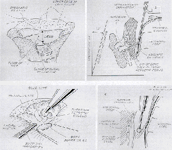
The retrolabirinthine approach was first described by Hitselberger and Pulec in 1972. It is performed through the mastoid air cells, with elevation of a dural flap between the labyrinth and the sigmoid sinus. The major advantage of this approach is that it provides direct access to the cerebellopontine angle without sacrificing hearing and without extensive cerebellar retraction. Its major disadvantage is the limited exposure that may be available when the mastoid air space is contracted.
Gherini in 1985 advocated the infralabirinthine approach. It is carried out by performing a simple mastoidectomy with preservation of the labyrinthine structures. The posterior fossa dura is exposed anteriorly to the sigmoid sinus, and work proceeds medially to the posterior semicircular canal and mastoid segment of the facial nerve. Dissection is carried inferiorly through the retrofacial air cells to the jugular bulb, which serves as the inferior limit to the dissection. The bulb may be skeletonized, however, and its superior portion may be carefully dissected to free it of its adjacent bony covering. It is then packed inferiorly with bone wax.
Maddox described in 1977 one-stage combined suboccipital translabirinthine approach that preserved the sigmoid sinus. House W and Hetselberger described the transcochlear approach to the cerebellopontine angle and clivus in 1976. This approach involves an extension of the translabirinthine approach anteriorly through drilling away the bone of the cochlea and exposing the dura over the petrous pyramis anteriorly to the internal auditory canal, Disadvantages of this procedure include the necessity for removing the facial nerve from the fallopian canal and transposing it posteriorly. The transotic approach is a modification advocated by Fisch U. and Gantz in which the posterior bony ear canal is removes and the facial nerve is not transposed. Fisch U. first described the infratmporal approach in 1977. Retraction or resection of the ramus of the mandible is accomplished to allow removal of bone over the petrous portion of the internal carotid artery.
The major disadvantages of this approach are the necessity for transposition of the facial nerve and hearing loss. Middle fossa-infratmporal fossa approach was proposed by Close et al. 1985 and Gale Gardner et al. 1985.
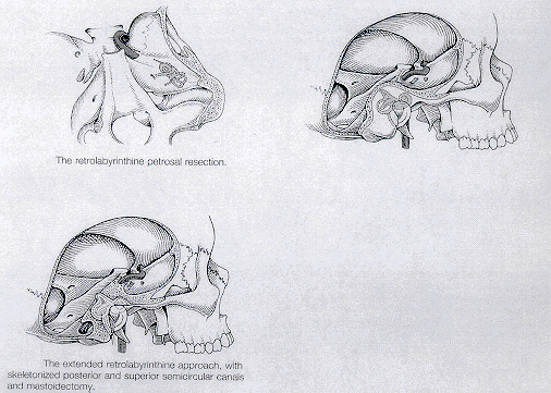
According to Koos W.T., Spetzler R.F. and Lang J 1993, the combined supratentorial and infratentorial approach is recommended for any tumour, that is situated above and below the tentorium along the petrous ridge, the clivus, or both. The relationship between the vascular structures is so important, that the surgeon who is unfamiliar with this region is urged to dissect several cadavers before attempting surgery. The petrous bone is drilled initially, exposing the dura and the sigmoid sinus. The extent of petrous bone resection depends on the desired exposure. The resection, and therefore the exposure, can be extensive even if hearing needs to be preserved. If, however, the entire inner ear can be sacrificed, the petrous bone resection can be maximized after mobilizing the seventh cranial nerve, thereby gaining a generous view of the base of the skull. When this approach is combined with the subtemporal and posterior fossa approach, the entire extent of the base of the skull can be visualized from the foramen magnum to the tip of the temporal fossa. If the facial nerve is completely drilled out of its canal, facial paresis that can persist for 6-12 months must be anticipated. If slightly less exposure is adequate, the seventh cranial nerve can be protected with a rim of bone and left in its normal anatomical course, to avoid facial palsy. The combined approach has been divided into three variations. The first is the retrolabirinthine exposure, which maintains the labyrinth intact, during the drilling of the petrous ridge, thus preserving hearing. The second is the translabirinthine exposure, in which the labyrinthine is removed and ipslateral hearing is thereby sacrificed. The third, and most extensive, approach is the transcochlear, in which the entire cochlea and the remainder of the petrous pyramid are sacrificed. The seventh cranial nerve is severed from its superficial petrosal branch and transposed from its canal.
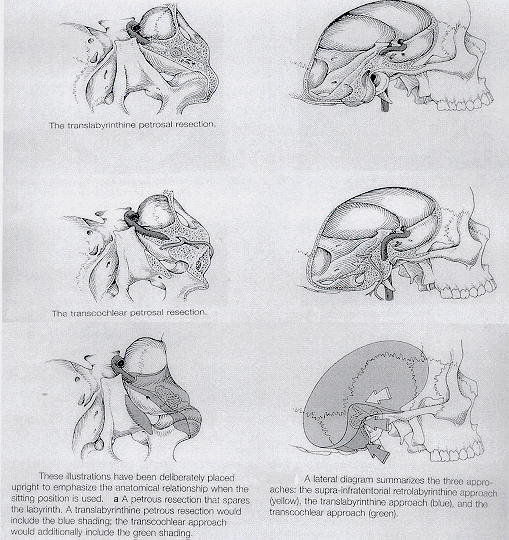
Tarlov E.C. divided tumours arising from the clivus into four groups. Tumours involving the dorsum sellae should be approached beneath the temporofrontal junction. Tumours involving the upper portion of the clivus can be best exposed subtemporally if a major or complete resection appears feasible preoperatively. The petrosal approach, drilling away the lateral portion of the petrous bone extramurally as described by AI-Mefty, permits a wide exposure of the upper clivus. Tumours of the lower clivus are best reached from a lateral approach drilling away the lateral portion of the foramen magnum and the first cervical vertebra and mobilizing the vertebral artery posteriorly.
For lesions of the midclivus, either the lateral suboccipital or the subtemporal approach is appropriate. A combined supratentorial and infratentorial approach to the clivus provides a wide exposure of this region without interfering with important draining veins. A combined temporal and suboccipital flap is turned down across the lateral sinus. The lateral sinus is legated anterior to the vein of Labbe. The tentorium, lateral sinus, and temporooccipital junction then are elevated, after the tentorium has been divided medial to the legated portion of the lateral sinus by extending the excision all the way through the tentorial hiatus.
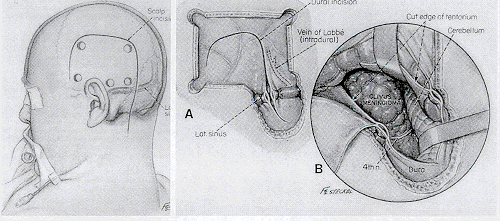
According to Schmidek H.H., the combined supratentorial and infratentorial approach is performed after exposure of the occipital bone through a lateral skin incision. An occipital craniotomy is then performed above the transverse sinus, after which the bone over the transverse sinus is thinned with a high-speed drill and removed. If the main reason for exposing the tentorium is to be able to obliterate the tumour's blood supply, ligating the transverse sinus may not be necessary. In this case, the dural incisions are made above and below the sinus, and the superior dural flap is situated lateral to the vein of Labbe. This method leaves a bridge of dura containing the transverse sinus. Superomedial retraction of the occipital lobe exposes the superior surface of the tentorium. Bridging veins from the cerebellar surface to the dura are cauterized and divided, thereby exposing the inferior surface of the tentorium. Assessing the characteristics of the tumour mass and interrupting its arterial feeders, should be possible. The tumour mass below the tentorium is often debulked first. If additional exposure is needed, the tentorium is split, avoiding the deep veins, posterior cerebral artery, and cranial nerves 111. IV and VI at the incisura. Even greater exposure is provided by ligation and splitting of the transverse sinus (Sato 0). Masses within the cerebellopontine angle are approached with the patient in either a sitting or prone position.
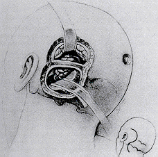
With the exception of very large tumours, these lesions can generally be managed through a laterally situated craniectomy. The skin incision is a long, S-shaped incision 2 cm medial to the mastoid process. The incision may extend high enough to allow it to be used to place an occipital burr hole. The craniectomy extends from the lower edge of the transverse sinus to the foramen magnum, from the medial edge of the sigmoid sinus about one half of the distance to the midline of the occiput. The mastoid air cells should occluded with bone wax. A single burr hole is then placed and the remaining bone thinned with a high-speed air drill before its removal with rongours.
References :
1. Gardner G. et al. Transtemporal approaches to the posterior cranial fossa . Operative Neurosurgical Techniques Henry H.Schmidek and William H.Sweet. Third Edition. 1995. W.B.Saunders Company. 843-850.
2. Gardner G. et al. Transtemporal approaches to the posterior cranial fossa. Am.J.Otol 6(Suppl): 1 18,1985
House W.F. et. al. The middle cranial fossa approach to lesions of the temporal bone and cerebellopontine angle. Neurosurgery. Robert H.Wilkins , Setti S. Rengachary Second Edition. 1996. McGraw-Hill.
I I 0 1 - 1 106.
3. John T. McElveen, Jr. The translabirinthine approach to cerebellopontine angle tumours. Neurosurgery. Robert
H.Wilkins , Setti S. Rengachary Second Edition. 1996. McGraw-Hill. I 1 07-1113.
4. King T.T., O'Connor: Translabirinthine approach to acoustic neurinoma. Operative Neurosurgical Techniques Henry
H.Schmidek and William H.Sweet. Third Edition. 1995. W.B.Saunders Company. 813-827.
5. Ojemann R.G. Suboccipital transmeatal approach to vestibular schwannoma . Operative Neurosurgical Techniques
Henry H.Schmidek and William H.Sweet. Third Edition. 1995. W.B.Saunders Company. 829-841.
6. Sato 0 : Transoccipital transtentorial approach for removal of cerebellar haemangioblastoma. Acta Neurochir (Wien) 59:195, 1981
7. Schmidek H.H. Surgical management of cerebellar tumours in adults . Operative Neurosurgical Techniques Henry H.Schmidek and William H.Sweet. Third Edition. 1995. W.B.Saunders Company. 791-800.
8. Tarlov E.C. Surgical management of tumours of the tentorium and clivus. Operative Neurosurgical Techniques Henry H.Schmidek and William H.Sweet. Third Edition. 1995. W.B.Saunders Company. 783-788.
9. Fisch U& Douglas Mattox. Microsurgery of the Skull Base.. 1988. George Thieme Verlage Stuttgart. New York.
10. Fisch U. : Infratemporal fossa approach for extensive tumours of the temporal bone and the base of the skull, in Siverstein H, Norrell H (eds): Neurological Surgery of the Ear. Birmingham, AL, Aesculapius, 1977, p 34.