Most of the site will reflect the ongoing surgical activity of Prof. Munir Elias MD., PhD. with brief slides and weekly activity. For reference to the academic and theoretical part, you are welcome to visit
neurosurgery.tv
Inomed Stockert Neuro N50. A versatile
RF lesion generator and stimulator for
countless applications and many uses
Multigen RF lesion generator .
14-SEPTEMBER-2014 ABDALLA WAFIQ FAHMAWI 28 YEARS
PERSISTENT CSF LEAK FROM THE LEFT MIDDLE FOSSA AFTER TRAUMA 10 YEARS AGO.
Anamnesis
The patient came to the clinic 19-July-2014
complaining of CSF leak after trauma 10 years
ago with recurrent episodes of meningitis for 5
years. Remission took place, but the last 2
months got CSF leak from the left nostril.
CT-scan done 19-June-2014 bad quality, not
informative.
On examination; the patient is neurologically
free, except the episodes of the CSF leak with
headache and decreased hearing left ear. The patient was sent for thorough
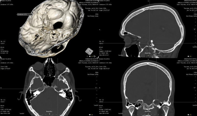
investigations: MRI 22-July-2014 showing CSF
pocket in the eroding left middle fossa. CT-scan
with with reconstruction using ORS Visual
showing the bony destruction reaching the
cochlea of the left pyramid.
Osteoplastic craniotomy immediately above the
left ear with reflection of the flap anterior.
Extradural approach to the subtemporal area. The
old fracture of the anterior part of the petrous
bone exposed. It was necessary to perform also
intradural approach to inspect the structures
intradurally. There was a huge bony defect in
the posterior part of the left cavernous sinus.
The left oculomotor nerve was seen running under
the defect. There was a huge fossa anterior to
the foramen ovale, through which the left V1 was
running. The dural defect was filled with muscle
harvested from the temporal muscle and glue used
to keep the muscle in place. This was done to
avoid mechanical trauma to the running left
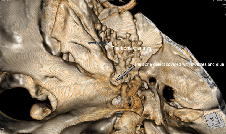
oculomotor nerve running under. Using bone
granules 5 ml the cavity anterior to the foramen
ovale and the bone defect in the petrous bone
were filled snuggly extradurally to prevent any
other unseen defects. Glue was applied over the
bone granules. The dura was closed water-tightly
and routine closure of the wound.
Smooth postoperative recovery.
The patient was sent to the ICU for 12 hours
observation.
Comments
The patient has CSF leak from the old
bony fracture of the left middle ear reaching the Eustachian
tube and eroding the entire left middle fossa. All the
suspected sources of the CSF leak must be dealt accordingly.
Skyra MRI with all clinical applications in the run since 28-Novemeber-2013.
Leica HM500
The World's first and the only Headmounted Microscope.
Freedom combined with Outstanding Vision, but very bad video recording and
documentation.
After long years TRUMPF TruSystem 7500 is running with in the neurosuite at
Shmaisani hospital starting from 23-March-2014
Inomed MER system
The bone defect in the tip of the pyramidal bone just anterior to
the left cochlea.
The bone defect covered by a piece of muscle with glue and bone
granules with glue to fill most of the gaps. The CT-scan done the
next day after surgery to confirm the taken actions are proper.
Notice: Not all operative activities
can be recorded due to lack of time.
Notice: Head injuries and very urgent surgeries are also
escaped from the plan .