Most of the site will reflect the ongoing surgical activity of Prof. Munir Elias MD., PhD. with brief slides and weekly activity. For reference to the academic and theoretical part, you are welcome to visit
neurosurgery.tv
Inomed Stockert Neuro N50. A versatile
RF lesion generator and stimulator for
countless applications and many uses
Multigen RF lesion generator .
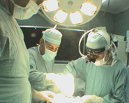
04-AUGUST-2009 ABDEL SALAM HASAN MUHAMED 15 YEARS
POST-TRAUMATIC HUGE BONY DEFECT IN THE LEFT FRONTO-TEMPORAL REGION.
Anamnesis:
The patient came to the
clinic 24-September-2008 after suffering severe
head injury 11-July-2008 and was unconscious for
more than 2 weeks. The patient underwent
decompressive craniotomy in the left
fronto-temporal region and the bone flap was
preserved in the refrigerator.
Serial CT-scans were
performed demonstrating the huge prolapse of the
brain from the bone defect.
On examination: the
patient had dense right
sided plegia with
hypalgesia of the right
side of the body with
spastic pattern. He
could walk with aid with
speech disturbances.
The father was advised
not to undergo surgery
for closing the bone
defect since the brain
still prolapsing with
severe degree and there
is no control of
epilepsy, for what he
was receiving Convulex
300 mg tid.
The patient was given
medications to decrease
the brain prolapse and
to rescue the still
surviving neural tissues
in the massive gliotic
left cerebral
hemisphere.
The patient was seen
05-October-2008 with MRI
of the brain
demonstrating the above
mentioned data.
The patient then came
07-July-2009 with slight
improvement of the right
plegia-paresis. The
patient is left handed
and the speech
normalized. There is no
prolapse of the brain
and the bone defect is
palpable with brain is
lax transmitting the
cardio-pulmonary
pulsation.
The patient was admitted
10 days ago, but he had
flue for what his
operation was postponed.
The removed bone was
requested from the other
hospital and inspected
and was autoclaved in
134 degrees for 15 min.
The old incision over
the left fronto-temporal
region was refreshed and
the dura was separated
from the the scalp. No
CSF leak was noted. The
left lateral ventricle
was punctured to rule
out the presence of
cystic cavities with any
brownish liquid
collection. A clear CSF
came out and the brain
came more relax.
The bone graft was
another time inspected
and cleaned meticulously
from debris and it
was fitting for the 90%
of the posterior area of
the defect. There were
chips of his bones in
the lower temporal
region not fitting with
construct, which were
removed and there were
inserted anterior to
fixed bone graft, where
there is still linear
bony defect. This
act helped in 2 points:
1. The removed bony
elements helped to
achieve more fitting of
the graft. 2. In case of
postoperative
complication, the only
suspected triggering
factor will be only the
graft, which was
preserved in another
hospital.
Routine closure of the wound
and smooth postoperative recovery.
Comments
In case that decompressive
laminectomy was performed, it is better to wait
until the brain becoming relax and not
protruding through the bone defect. It needs at
least 6-12 months depending at the severity of
injury and the presence of other triggering
factors and the epileptic activity.
The methods of bone
preservation are deferent and in this case
freezing the bone was performed. The shape and
texture of the bone was acceptable for what it
was used to cover the bone defect. To avoid
possible contamination, it is preferable to
autoclave the bone graft at 134 degrees for 15
min.
In skull X-rays the bone
usually is translucent and becoming visible
several months after surgery.
Surge of calcium could have
place in the postoperative period, for what Ca
level must be recorded for several days.
Please! wait for 3-5 min till the
video start to load. It depends upon the internet
connection.
Notice: Not all operative activities
can be recorded due to lack of time.
Notice: Head injuries and very urgent surgeries are also
escaped from the plan .