|
The patient came to the clinic
23-February-2006 complaining of weak right UL with numbness of the
right median nerve distribution. The patient suffered RTA
16-October-2005 without LOC, but he immediately started to complain
of the above mentioned. He is a known diabetic for 5
years.
MRI done 28-November-2005 showing PCD C5-6
with wedge fracture of C5. Repeat MRI done 16-February-2006 showing
deterioration of the condition with PCD C5-6. C6-7 with the
cervical X-rays demonstrating overmobility of C3 down to C7.
On examination: The patient had neck pain when
looking to right up and down with weak grip, extension of the
right hand and right triceps muscle with hypalgesia of the second
and third finger right hand. The patient was sent for further
studies and PCD C4-5 was also confirmed and wedging of C4 was noted.
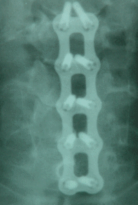
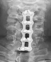
The patient was operated. Discectomy C4-5, 5-6,
6-7 was done with removal of the right extrusion at the right side
of C6-7. Using Stryker reflex hybrid ACP system, fusion of C3 down
with C7 was performed, using four level fixation.
Immediate postoperative recovery was uneventful,
but the patient progressed severe oedema at the site of the surgery
with and sent for ICU care 03-March-2006 at 1.00 p.m. and given 2
units human albumin. No surgical emphysema and laryngeal oedema is
the predominant picture.
The patient's condition deteriorated and he was
taken at 3.00 p.m. to the operating room. During intubation the
epiglottis and the surrounding tissues were swollen. Exploration of
the wound revealed moderate hematoma overlying the construct
about 7-80 ml thick in consistency without active bleeding. It was
removed and all the seen veins coagulated. Inspection of the
esophagus and the trachea for possible tears were negative. The
carotid sheath was intact and the thyroid also.
The wound was washed with saline and gentamicin
and ready-vac drain No 10 was inserted and the patient left in
ventilator to the next day. 1 unit blood and 6 units FFP were given
for the possible unrecognized coagulopathy.
The next morning 04-March-2006 the patient
progressed left sided pneumothorax for what UWS was applied to the
left side. CT-scan performed at 9.30 a.m. showing no haematoma at
the operative site and mild bilateral heamothorax. It was decided to
keep the patient in ventilation for further 2 days.
The patient was extubated after 2 days and the
chest tube removed the next day and the patient was transferred
to the ward 8-March-2006 and discharged in good condition with
improved neurologic condition 12-March-2006.
Comments:
1. The patient got rupture of the OPLL at all the
mentioned levels due to severe hyperflexion injury. This was the
cause of his ruptured disci and overmobility of all these segments.
It was possible to find the site of the ruptures.
2. Conservative treatment is unlikely will
resolve his problem, and surgical decompression and fixation, put
the patient in the safe side from developing myelopathic syndrome.
3. It is the fist time in my 26 years of personal
experience seeing a case with a slowly progressive hematoma
progressing to that degree, that evacuation of the hematoma was
needed and for extreme precaution putting him in sedation with
muscle relaxants after such surgery. The most possible cause of the
haematoma was a torn small vein , which was silent during surgery ,
but escalated and gradual enlargement of the oozing mass
several hours after the operation.
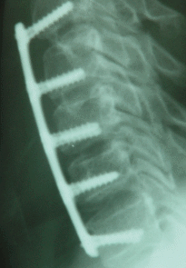
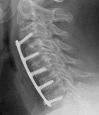
4. Tow signs must be focused to attention: The
patient was unable to breath in the supine position, for what
he refused the CT-scan. This could be explained retrospectively,
that when the patient extend his head, the trachea suffer more
compression from behind. If you pay attention to the below picture
in lateral view, you can see some distance between the construct and
the trachea, which was in evolution at that time.
5. When inserting a such large device, a huge
traction is needed. It is preferable to coagulate and sharp cut all
the running small veins in the route and at construct site, so as to
avoid such complication.
|