Most of the site will reflect the ongoing surgical activity of Prof. Munir Elias MD., PhD. with brief slides and weekly activity. For reference to the academic and theoretical part, you are welcome to visit
neurosurgery.tv
Inomed Stockert Neuro N50. A versatile
RF lesion generator and stimulator for
countless applications and many uses
Multigen RF lesion generator .
24-NOVEMBER-2011 AYSHA SULAYMAN AHMAD 24 YEARS
PRESACRAL GIANT MASS WITH ANOTHER MASS ORIGINATING FROM THE RIGHT LUMBO-SACRAL
PLEXUS.
Anamnesis
The
patient came to the clinic 19-November-2011
complaining of constipation, urgency and
frequency with strange feeling when setting. The
patient was operated 02-June-2010 for pelvic
mass through laparoscopic approach, but the
patient did not feel any improvement. CT-scan of
the pelvis done 28-May-2010 before surgery
showing huge presacral mass 97x117 mm in
dimensions. There is no histologic study, nor
postoperative control studies.
MRI of the
pelvis done 17-November-2011 showing the same
previous mass and a separate mass in the region
of the right lumbo-sacral plexus. They are well
separated, but adherent to each other.
On
examination: There is weak
dorsi and planterflexion right foot 4/5.
The
patient was sent for MRI of the lumbo-sacral
spine and CT-scan of the pelvis, which were done
20-Noveember-2011 ruling out any CSF connection
between the masses and the intradural structures
of the spinal cord.
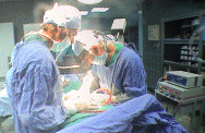
Right pelvic rim extraperitoneal approach with
extension medially through part of the old
caesarian incision. The running lateral femoral
cutaneous nerve was dissected and preserved. The
dissection was taken over the iliac and psoas
muscle. The femoral nerve was identified and was
checked with ISIS Inomed HighLine 32 channel
Neurosxplorer neuronavigation. The external
iliac artery and vein were identified and
preserved. The psoas muscle was retracted
lateral to expose the obturator nerve. The
internal iliac vein with the running below the
L4 and L5 roots were identified and check with
electrophysiological navigator. There is no
tumor there and there is adhesion due to old
inflammatory process. More medial
dissection was carried out, but no proper
cleavage was seen to resect the huge cyst, which
had thick capsule, which was punctured and
around 150 yellow-brown thick fluid was obtained
and sent for histologic and bacteriologic
studies. A cysto-fix was inserted in the
coccygeal area anterior to the sacrum and around
400 same color puss was evacuated. 150 ml
Renografin was diluted with 500 ml normal saline
was used to irrigate the cyst, which was studied
using C-arm. The cystic mass could be
identified after filling the cavity with with
400 ml of the mentioned solution. The wall of
the cyst was stuck to all anatomical structures
and it was impossible to separate it. Some parts
of the capsule was sent for histologic studies
and swollen lymphnodes also. The cavity then was
irrigated with normal saline and complete
evacuation of the cyst was achieved and checked
by the C-arm. A drain was kept to big cyst and
the wound was closed by layers and Ready-vac
drain was put under the skin lateral to the
wound.
Smooth postoperative
recovery with improvement of the power of
the right foot.
Please! wait for 3-5 min till the
video start to load. It depends upon the internet
connection.
Comments
The patient was operated 17 months ago and
laparoscopic intervention was done. These 2
masses cannot be dealt by this approach.
The smaller mass originating from the neural
structures is more important to explore and
remove than evacuation the huge cyst.
Retrospectively, it was wise to evacuate the
cyst through coccygeal route and inject the die
and do studies to evaluate the multilobular
cyst. But even when doing this, the neurosurgeon
will remain unsure about the mass in the right
lumbosacral area until he do proper exploration
of this area.
Follow Up
The final histologic result was scarococcygeal
teratoma with no malignant cells. Other
elements of germ cell tumour cannot be excluded.
All investigations for tbc were negative and all
laboratory findings were not specific.
Notice: Not all operative activities
can be recorded due to lack of time.
Notice: Head injuries and very urgent surgeries are also
escaped from the plan .