Inomed Stockert Neuro N50. A versatile
RF lesion generator and stimulator for
countless applications and many uses
Multigen RF lesion generator .
03-DECEMBER-2019 AMAL MIDHAT ABDEL-QADER 64 YEARS
HUGE MID THIRD MENINGIOMA WITH EXTENSION TO BOTH PARIETAL CONVEXITIES MORE TO
LEFT AND INVOLVEMENT OF THE FALX CEREBRI AND INSIDE THE SSS.
Anamnesis
The patient came to the clinic 09-November-2019
complaining inability to walk the last 3 months
with epi attacks. MRI of the brain bad quality,
not complete study performed 27-September-2019
showing lesion left parieto-frontal and less in
the right.
On examination: there is weak grip right hand
4/5, extensors right hand and right biceps 3/5.
Weak dorsi and planterflexion right foot 3/5,
abduction right knee 4/5, right quadriceps
femoris and right iliopsoas muscles 2/5.
The patient was sent for investigations
with complete protocol of MRI with clinical
applications, including fMRI, which were done
12-November-2019 showing huge meningioma
occupying both parietal areas more the left with
involvement of the falx cerebri and tumor
was extending inside the mid third of the SSS
with draining veins anterior and posterior to
the involved SSS. The motor area was located
posterior to the lesion. The patient was sent for
cardio evaluation.
In semi-setting position with
vertex is at its superior position, trying to
decrease the pressure of the SSS, extended wide
craniotomy of both fronto-parietal area with
reflexion of the flap to the left, trying during
that the avoid traction injury to the SSS. The
tumorous bone flap sent for sterilization to kill
the inside growing tumor. The dura was opened
lateral to the left meningioma margin and followed
to the edge of the SS anterior and posterior. The
tumor was rubbery in consistency and highly
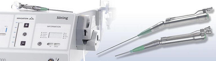
vascular. Using SONOCA 300 with different handles
the tumor was resected step-wise. Part of it was
sent for histologic investigations. The resection
was carried down until the falx cerebri was seen.
Opening the right side confirmed that there is no
apparent tumor and the presence of huge draining
veins and Pacchoinian granulation, restricted the
full exposure of the area to explore the other side
of the right wall of the SSS, which in fact was full
of tumor. The dura in the right side was closed and
the patient was sent for intraoperative MRI
investigation with contrast and MRV. The defect of
the SSS still the same and the tumor is inside and
around the SSS. After consulting all the team
members, it was decided to satisfy this degree of
resection and to be followed by radiation 8-10
months after surgery. The sterilized bone was
returned back. Routine closure of the wound.
Smooth postoperative recovery
with right severe spastic paraparesis. She was sent to the
ICU.
Follow Up
The patient 4 hours after surgery in the ICU is
alert responding to verbal stimuli talking and
the paresis regressed dramatically.
The final histologic result was that of
meningioma, transitional type (mixed
meningothelial and fibrous); WHO Grade I. (Prof.
Yahia F. Dajani 07-12-2019).
The patient came three times to the clinic with
dramatic improvement of her neurologic status to
evacuate fluid collection over the bone flap.
Comments
This case is challenging with its
involvement of the SSS. Without violating it the patient
progressed immediate postoperative deep right sided spastic
paraparesis.
What if the SSS was violated? Death could
be one of the options which is not desirable in account.
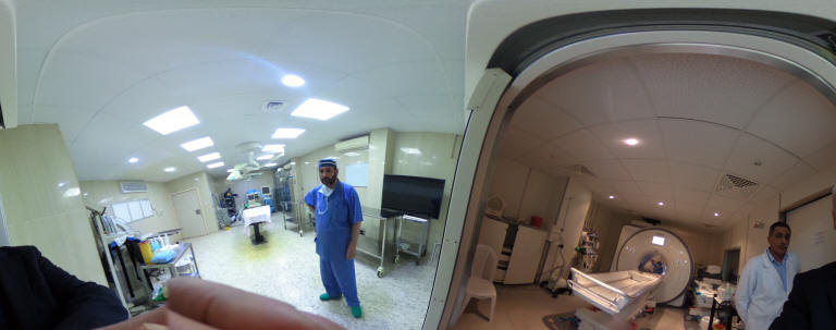
Skyra MRI with all clinical applications in the run since 28-Novemeber-2013.
Inomed Riechert-Mundinger System, with three point
fixation is the most accurate system in the market. The microdrive and
its sensor gives feed back about the localization.
Inomed MER system
Leica HM500
The World's first and the only Head mounted Microscope.
Freedom combined with Outstanding Vision, but very bad video recording and
documentation.
After long years TRUMPF TruSystem 7500 is running with in the neurosuite at
Shmaisani hospital starting from 23-March-2014
SONOCA 300
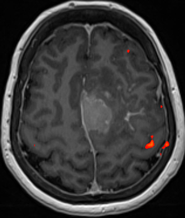
Fig-1: fMRI of the right hand tapping.
Notice: Not all operative activities
can be recorded due to lack of time.
Notice: Head injuries and very urgent surgeries are also
escaped from the plan .