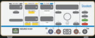
Inomed Stockert Neuro N50. A versatile
RF lesion generator and stimulator for
countless applications and many uses
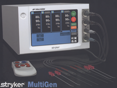
Multigen RF lesion generator .
09-JUNE-2026 AMIRA MAHER AL-SHAREEF
28 YEARS INSERTION OF THE MEDTRONIC SHUNT FROM THE EXTERNAL DRAIN.
Anamnesis
The patient was operated by me
2 days
ago for revision of the craniotomy site and
insertion of the external drain. The CSF coming
to the external drain is permitting insertion of
the Medtronic medium pressure to the abdominal
cavity.
Skin incision was made behind
the craniotomy site and the intracranial tube
was withdrawn around 4 cm to keep the
proximal end away from the pineal gland. The
medium pressure Medtronic valve with the
abdominal part were inserted to the left side of
the abdomen.
The patient showed smooth
recovery and sent to the ward.
FOLLOW UP
The patient then came to
Emergency 17-June-2026 at 7.30 p.m. due to
convulsions which terminated before arrival to
the hospital. The patient in bed talking and
having verbal contact. All the stitches were
removed and and the valve is functioning.
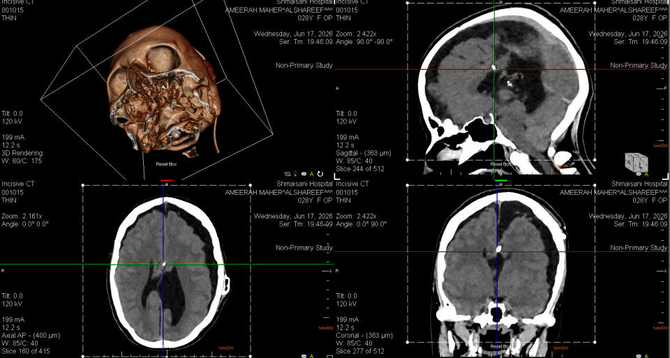
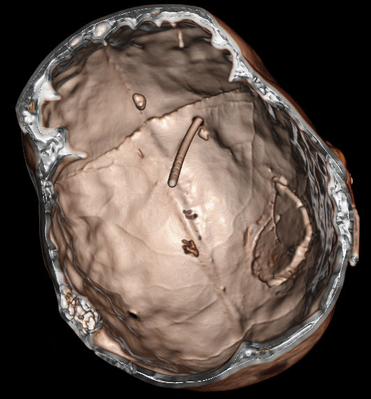
CT-scan of the brain was done, which showed
acceptable location of the shunt, but shrinking
of both cerebral hemispheres more the left with
slit upper III ventricle with cerebellar
hemisphere pushed down and the previous tonsilar
herniation took place and the prepontine space
is lacking CSF space. The patient sent home and
a plan was explained.
The patient then came 12.00
p.m. and the patient was dead for at least 15
min.
Comments
The case is very complicated, because
there are no previous MRI data to compare with.
The VPS has small reservoir, and not
functioning for unknown period.
The family have controversial
information, that the surgeon cannot configure what is going
on, but the Jacksonian attack of the right upper limb, which
took place 2 years ago and the the drop right foot, can
denote that the seizure activity took place from the leg and
hand area of the sensomotor strip left hemisphere.
This case make it clear to not reach the
pineal body and to keep at least 15 mm above the target to
avoid shifting of the brain after decompression and upward
migration of the brain stem.
Do not leave loops outside the bur hole
with no proper fixation to prevent slippage of the tube
intracranially.
During insertion of the shunt the
intracranial part was withdrawn 4 cm to keep away from the
pineal body region.
The epiactivity was diencephalic mixed
with secondary generalized tonic clonic.
The cause of death still unknown. but
could be due events happening at the respiratory cardiac
centers of the pons such as dorsal Respiratory Group (DRG):
Primarily located within the nucleus tractus solitarius (NTS).
It functions as an inspiratory center, receiving sensory
input (such as lung stretch and blood chemistry) and
triggering the nerves (like the phrenic nerve) that cause
the diaphragm to contract.
Ventral Respiratory Group (VRG): Located
ventrally in the medulla. It contains both inspiratory and
expiratory neurons and controls forced exhalation and
accessory muscles. Pre-Bötzinger Complex: Found within the
VRG, this specific region acts as the central pacemaker,
generating the fundamental, rhythmic electrical drive for
breathing. Nucleus Ambiguus and Nucleus Retroambigualis:
These nuclei provide motor control for the upper airway
muscles (larynx, pharynx, and soft palate) and contribute to
both inspiration and expiration. 2. Pontine Respiratory
Group (PRG) Located in the pons, this group fine-tunes the
rhythm generated by the medulla to ensure smooth, controlled
breathing.
Pneumotaxic Center: Associated with the
parabrachial nucleus and the Kölliker-Fuse nucleus. It
actively inhibits the inspiratory phase, acting as an
"off-switch" to limit inspiration, thereby controlling the
depth and rate of breathing. Apneustic Center: Located in
the lower pons, it stimulates inspiratory neurons to prolong
inhalation. It is normally suppressed by the pneumotaxic.
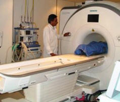
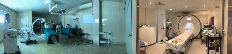
Skyra MRI with all clinical applications in the run since 28-Novemeber-2013.
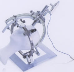
Inomed Riechert-Mundinger System, with three point
fixation is the most accurate system in the market. The microdrive and
its sensor gives feed back about the localization.
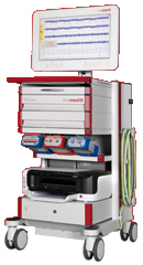
Inomed MER system
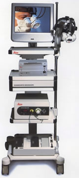
Leica HM500
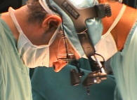
The World's first and the only Head mounted Microscope.
Freedom combined with Outstanding Vision, but very bad video recording and
documentation.
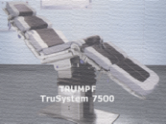
After long years TRUMPF TruSystem 7500 is running with in the neurosuite at
Shmaisani hospital starting from 23-March-2014
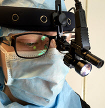
LooksCam II Xenosys in the run starting from 14-March-2021 with
SheerVision TTL x4 magnification.
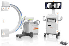
Cios-Spin flat panel in the run.
CT-scan done the last admission showing the shunt and deferent
changes of the brain and brain-stem. 17-June-2026 at 8.00 p.m.
Notice: Not all operative activities
can be recorded due to lack of time.
Notice: Head injuries and very urgent surgeries are also
escaped from the plan .