|
The patient was brought to ICU in semiconscious state with
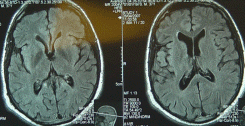
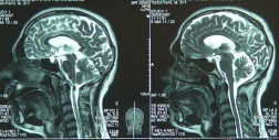
progressive deterioration within 2 days. MRI performed today showing
colloid cyst with acute hydrocephalus. Mannitol with decadron
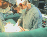
started and within 3-4 hours the patient was operated. Right
tranfrontal transcortical approach to the right lateral ventricle.
The chocolate fluid was evacuated through syringe without
contaminating the CSF. After that, the ventricles became lax and it
was possible to see the floor of the third ventricle. The cyst had
multicompartment structure, for what it was dealt with caution.
The wall of the cyst was highly vascular and stuck with choroid
plexus and many arterial feeders were coagulated and bisected. Part
of the cyst wall was the thalamic vein, which was preserved to avoid
venous occlusion.
After complete separation of the wall of the colloid cyst it was
radically removed. An external drain was inserted to the right
lateral ventricle and routine closure of the wound.
Smooth
postoperative recovery and the patient was completely alert and
cooperative the next day with no CSF coming through the external
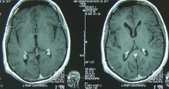
drain. CT-scan done the morning of 08-May-2006 showing complete
removal of the mass with air still in the lateral ventricle and the
subfrontal area. The patient showed dramatic recovery and the
medications were tapered within several days and discharged
15-May-2006. The final histologic result confirmed the presence of
colloid cyst. For theoretical references concerning colloid cysts,
click here! |