|
The patient came came to the clinic 11-June-2006 complaining of
LBP for 7 months with bilateral sciatica more the left. She
was limping with scoliotic stance. SLRS was 60 degrees in the right
and 40 degrees in the left. She had hypalgesia of the left S1 root
with weak dorsiflexion both feet. MRI performed, showing LCS L4-5
with huge extrusion at the right side. The patient was operated:
bilateral flavotomy with foraminotomy to both L5 roots was done. The
extrusion was removed from the right side in one piece. Seeking the
defect in the annulus fibrosis showed very small defect, for what it
was intentionally decided not to violate the intradiscal space.
Routine closure with smooth postoperative recovery.
Comments:
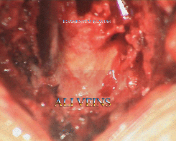
1. During drilling of the medial aspects of the facets, there is
a persistent vein in the upper corner of the facet, which I gave it
the name of Ali Bayati vein, the name of my lovely assistant. The
practical importance of this vein, that it indicate the limits of
the upper corner drilling limits, after what the ligamentum flavum
disappear and drilling must be performed with precaution, using some
device to protect the epidural fat or the dura from possible injury
during drilling.
 2. When LCS and relative huge extrusion took place
simultaneously, it is hard to tell which root/roots , most affected,
and it is hard to predict the localization of the extrusion. |