Inomed Stockert Neuro N50. A versatile
RF lesion generator and stimulator for
countless applications and many uses
Multigen RF lesion generator .
30-MARCH-2021 FATMEH AHMAD AL-RIYATI 45 YEARS
HUGE RECURRENCE OF MEDULLOBLASTOMA IN THE RIGHT FRONTAL AND SMALLER LOCAL.
Comments
The medulloblastoma in adults, despite
radical resection and radiotherapy can recur after 10-25
years as in this case.
The tumor at the posterior
fossa was ignored because it was not causing any
shift of the structures, in opposite to the
frontal huge mass which causing an impending
conning.
This frontal part of the
tumor could be a meningioma, because it has
matrix and the consistency is looking as
meningioma, but spectroscopy of the frontal part
was more malignant than the part in the
posterior fossa.
It turned to be meningioma
with malignant features, but total resection
with matrix, achieved the final goal.
Late radionecrosis of the
right cerebellar area must be considered in
mind.
Anamnesis
The patient was operated by me 05-April-1999 for
medulloblastoma and was doing fine and underwent
radiation 35 Gy 20 fractions whole body and 20
Gy 10 fractions over 6 weeks. The patient
came several times over the years and several
MRI of the brain ruled out recurrence. The
patient then came 19-August-2014 with MRI of the
brain showing small mass in the right frontal
lobe. The patient then was sent for
investigations, but she disappeared and came
10-September-2018 with numbness left upper limb
and both lower limbs for 1 year with fainting
attacks. The decreased hearing right side before
the first surgery. The patient was sent for
investigation. MRI brain done 10-September-2018
showing huge mass in the right frontal area
shifting the midline structures to the left.
The patient was advised to undergo surgery, but
she disappeared.
The husband by telephone, telling that she is
deteriorating for what she was admitted to
Shmaisani hospital 28-March-2021. The patient is
bedridden walking with help of 2 persons with
difficulty, hallucinating at times and with
difficult verbal communication and she could
remember me as her doctor. Left sided
hemihypalgesia and paresis.
The patient was sent for investigations and MRI
repeated 29-March-2021 under G.A. showing
considerable enlargement of the mass in the
right frontal area 6X6 cm in dimensions, pushing
down the brain stem, including the red nucleus.
There is appearance of recurrence at the
previous surgery in the posterior fossa but not
causing compression, not violating the 4th
ventricle, but reaching the upper vermis up the
edge of tentorium. Spectroscopy showed high
cholesterine level of the frontal part, alarming
malignant nature of this part. The mass in the
right cerebellar hemisphere is less malignant
with normal cholesterine level.
Due to preoperative medication, after one day,
the function of the reticular activating
formation improved and the patient became more
conscious to show full blown frontal lobe
syndrome with aggressiveness, for what
preoperative interrogation with the patient will
be impossible. Consent for surgery was obtained
from her husband from Saudia by telephone, due
to Covid restrictions.
Right frontal osteoplastic
craniotomy with reflection of the flap to the
right ear. The dura was suck to the bone, that
it was torn during opening. The tumor was seen,
as be having matrix to the dura at its medial
aspect. It was highly vascular, reddish
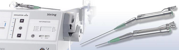
resembling a meningioma. Using SONOCA 300
ultrasonic aspirator and other facilities, piece
meal resection of the tumor was achieved. It was
multiple consistency, with rich vascularity. The
solid parts were impossible to remove by SONOCA
300.It was removed by pituitaries after
coagulation. The tumor margins were followed
until all its parts were removed. Several parts
of the tumor were sent for histological
verification. It was possible to see the right
olfactory tract running anterior and inferior.
The falx cerebri was at its medial border with
pericallosal arteries running above the corpus
callosum. Some tiny structures were preserved to
avoid spillage of CSF from the ventricles to
prevent contamination of the tumor to the
ventricular system. The bed of the tumor was
covered with three pieces of Surgicele to
control bleeding. The matrix of the tumor was
coagulated and the dural defect (which actually
the matrix of the tumor) was closed using
part of the muscle of the reflected flap to
achieve water-tight closure. Routine
closure of the wound. Before extubation, the patient
was sent to MRI and 2W axial MRI confirmed total
resection of the tumor.
Smooth postoperative recovery.
The power of the left side of the body improved.
She was sent to the ICU for 20 hours
observation.
Follow Up
The final histologic result was meningioma WHO
Grade II. (Prof. Yahia F. Dajani, M. B. Ch. B.
(Bristol), F. R. C. Path. ( London) Consultant
Pathologist 01/04/2021).
The patient started to show some regression of
the frontal lobe syndrome and can walk in the
corridor with clean wound.
After decreasing the Decadron dose, the
patient's frontal lobe syndrome became more
prominent and check CT-scan of the brain done
03-April-2021 was uneventful.
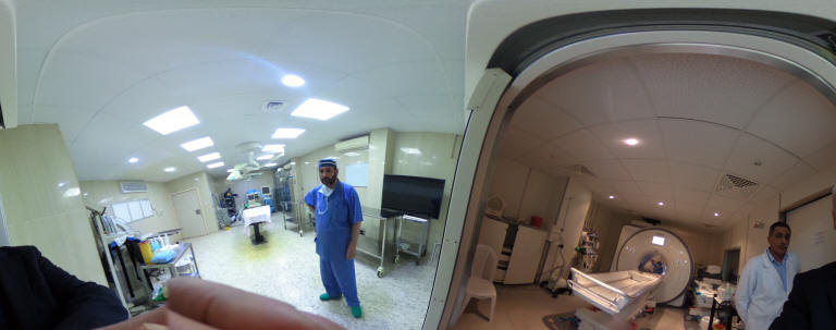
Skyra MRI with all clinical applications in the run since 28-Novemeber-2013.
Inomed Riechert-Mundinger System, with three point
fixation is the most accurate system in the market. The microdrive and
its sensor gives feed back about the localization.
Inomed MER system
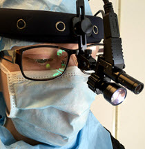
Leica HM500
The World's first and the only Head mounted Microscope.
Freedom combined with Outstanding Vision, but very bad video recording and
documentation.
After long years TRUMPF TruSystem 7500 is running with in the neurosuite at
Shmaisani hospital starting from 23-March-2014
LooksCam II Xenosys in the run starting from 14-March-2021 with
SheerVision TTL x4 magnification.
SONOCA 300
Notice: Not all operative activities
can be recorded due to lack of time.
Notice: Head injuries and very urgent surgeries are also
escaped from the plan .