Inomed Stockert Neuro N50. A versatile
RF lesion generator and stimulator for
countless applications and many uses
Multigen RF lesion generator .
27-NOVEMBER-2023 FATMEH TAHER AL-QAHEM 55 YEARS
INTRAMEDULLARY EPENDYMOMA EXTENDING FROM C5 DOWN TO D1 WITH TETRAPARESIS AND
PLEGIA BOTH FEET.
Anamnesis
The patient a Yemeni citizen came to the clinic 18-November-2023
complaining of difficult walking for 4 years
with more weakness of the left leg with gradual
deterioration. In wheelchair for 8 months. MRI
cervico-dorsal spine done 19-November-2023
showing intramedullary ependymoma extending from
C5 down to D2. MRI of the brain was normal.
On examination, the patient is in wheelchair
with weak both hands -4/5 both iliopsoas muscles
1/5 quadriceps femoris 3/5, abduction and knees
adduction 2/5 no dorsi or planterflexion both
feet. There is hypalgesia D3-D6 both sides and
hypalgesia down to the right leg. Micturition
and defecation preserved. Deep reflexes lower
limbs exaggerated and Babinski more brisk with
clonus in the left.
The patient was sent for investigations and MRI
cervical and dorsal spine done the same day
demonstrating the below figures.
Laminectomy of lower half of
C5 down to upper half of D2. The
dura was opened and reflected to the sides.
Inspection of the spinal cord with the violet
tumor transparently seen at the right upper half
of the field. Longitudinal incision above this
area without violating the capillaries. The
tumor was soft but not suckable, for what
piece-meal resection was applied. The spinal
cord was gently preserved. Practical total
removal of the tumor was achieved, A tiny
residual adherent to the vein was coagulated and
left to avoid trauma to the vein. The spinal
cord became lax and no palpable mass inside it.
Water-tight closure of the dura and routine
closure of the wound. The patient was put in Reverse Trendelenburg
position with Valsalva maneuver and
hyperventilation. No CSF leak. Before weaning
the patient intraoperative MRI was done. No
residual is seen. She was sent to the
ward.
FOLLOW UP
Too early now.
Comments
The patient has huge ependymoma with
plegia of the lower limbs and paresis of the hands. Surgery
is mandatory.
Even with practical removal of the tumor,
postoperative radiation is mandatory.
Skyra MRI with all clinical applications in the run since 28-Novemeber-2013.
Inomed Riechert-Mundinger System, with three point
fixation is the most accurate system in the market. The microdrive and
its sensor gives feed back about the localization.
Inomed MER system
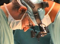
Leica HM500
The World's first and the only Head mounted Microscope.
Freedom combined with Outstanding Vision, but very bad video recording and
documentation.
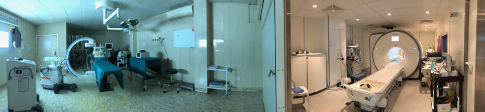
After long years TRUMPF TruSystem 7500 is running with in the neurosuite at
Shmaisani hospital starting from 23-March-2014
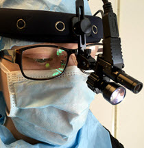
LooksCam II Xenosys in the run starting from 14-March-2021 with
SheerVision TTL x4 magnification.
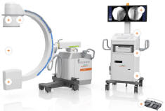
Cios-Spin flat panel in the run.
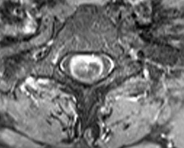
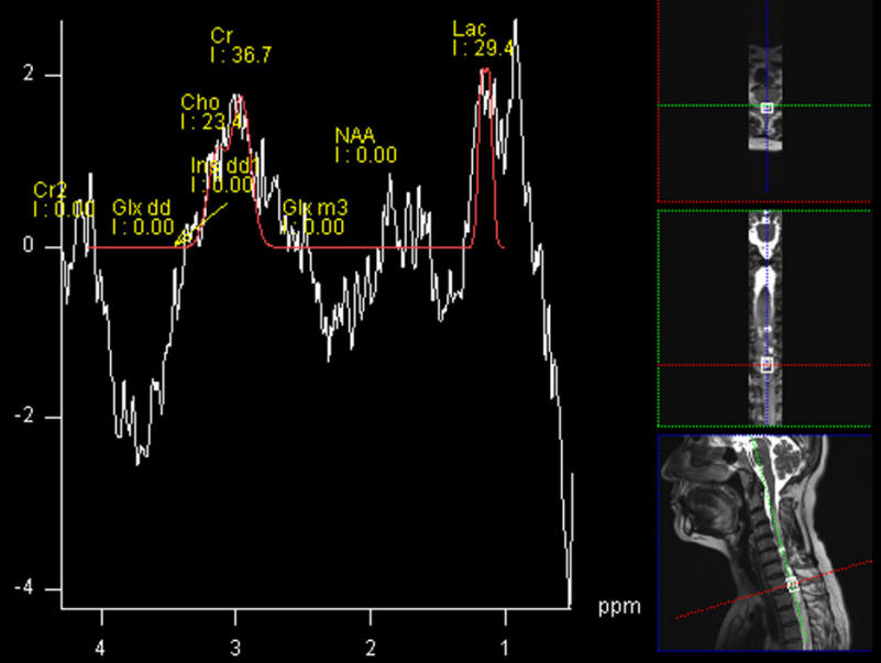
Fig-1: Axial view showing solid component surrounded by collection.
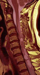
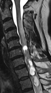
Fig-2: The ependymoma without contrast. Fig:-3 Saggital view with
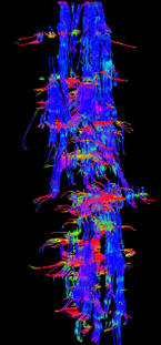
contrast. Fig:-4 Fibertraking confirming presence of fibers around
the tumor. Fig:-Spectroscopy ruling out malignant nature of the
lesion.
For more information about ependymomas,
click here.
Notice: Not all operative activities
can be recorded due to lack of time.
Notice: Head injuries and very urgent surgeries are also
escaped from the plan .