Inomed Stockert Neuro N50. A versatile
RF lesion generator and stimulator for
countless applications and many uses
Multigen RF lesion generator .
26-MAY-2025 GHAZWAN SAMI AMEEN 55
YEARS: SECOND PART OF THE SURGERY TO DECOMPRESS THE INVOLVED D7.8 BY
THE METASTATIC MALIGNANT PAPILLARY THYROID CARCINOMA AND STABILIZE
THE SPINE.
Anamnesis
The patient was operated by me
15-May-2025 and
the surgery was stopped due to massive bleeding
after 13 hours and sent to ICU to correct his
homeostasis.
On examination, the patient is improved for his
previous paraplegia, but fixation of the spine
did not performed and the tumor was removed
around 40% and the spinal cord was decompressed.
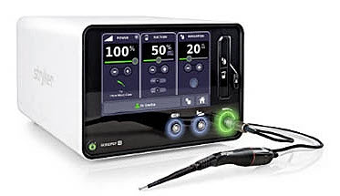
Sonopet IQ was prepared for the procedure.
With the patient in right
side position, the previous incision was
refreshed and the the incision over the left
chest cage extended more. It seems that
during the 11 days the tumor progressed in size
denoting the aggressive course. The tumorous
left D8-7 lateral mass was removed with the left
D7 was removed. The same was done to the
tumorous articulation of left D8-9 with removal
of the tumorous rib of D9. Using Sonopet
IQ was of no help in decompressing the
extrapleural with Barracuda tip, because the
tumor was firm solid and contained bones. The
tumor was decompressed by several means of
cauteries and bipolars and drills. The pleura
was opened and the real dimensions and
configuration of the mass was realized. This
step could yield to remove almost all the
tumor in the area keep the dissection
extrapleurally. The left lateral wall of D6 and
D9 were exposed and 2-0 angle end plates were
applied to the lateral walls of D6 and D9.
5.5x40 mm F.A.S Armada screws were inserted 2
screws to each bones. So as to accept the
Nuvasive X-core 2- 41-46 expandable corpectomy
cage, further drilling of D7 healthy bone was
done. The disc space of D8-9 was eaten by the
tumor. The construct was inserted and
distracted and rigid fixation obtained. We
used Attrax bone graft 10 cc was used to fill
the inside the corpectomy cage, because the ribs
and laminae were tumorous. 2 Rods slightly
bended to accept the lordotic area of the spine.
and left side fusion of the D6-D9 was achieved.
Hemostasis and insertion of underwater seal
inserted and the pleura was closed. Ready Vac
drain was also applied. Routine closure of the
wounds. The operation time was 24 hours, for
what the patient was put in ventilator for 24
hors.
Sonopet IQ in the run.
FOLLOW UP
In the ICU, blood transfusion, FFP was given and
all the homeostatic parameters was observed and
corrected accordingly. The patient the next day
28-May-2025 extubated and with no neurologic
deficits.
same protocol done 24-May-2025. Votive pleural effusion
took place both side more the left side.
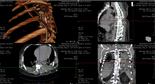
ORS Visual showing the removed tumor and inserted
the Nuvasive corpectomy device done 31-May-2025
In 08-June-2025, the patient was ready for
discharge after removing the UWS the day before
and repeating CXR which was acceptable, during
readiness for discharge, but the patient still
in the ICU, he progressed massive pulmonary
embolism and resuscitation failed and he died at
2.30 p.m.
Comments
The tumor was bleeding vigorously, for
what so as not to loose the patient, the surgery was
prepared promptly. and the surgical mission succeeded.
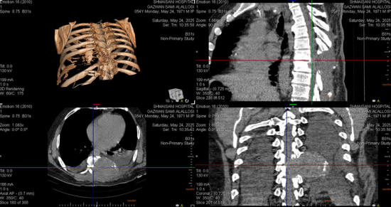
The patient performed chest CT-angio
after the first surgery and it was normal denying absence of
pulmonary embolism. There were no warning signs to predict
such event, which made the patient death.
The patient during this period received
more than 60 units of packed cells and 120 units FFP and 50
units platelets. At these circumstances, it is difficult
cover him with anticoagulants. It seems that even a minimal
amount of anticoagulants will increase the amount of
transfusion, but could avoid the pulmonary embolism.
In medicine, the puzzle is more complex
than chess or AI technology, and you cannot predict when the
catastrophe will take precedence.
Skyra MRI with all clinical applications in the run since 28-Novemeber-2013.
Inomed Riechert-Mundinger System, with three point
fixation is the most accurate system in the market. The microdrive and
its sensor gives feed back about the localization.
Inomed MER system
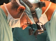
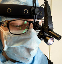
Leica HM500
The World's first and the only Head mounted Microscope.
Freedom combined with Outstanding Vision, but very bad video recording and
documentation.
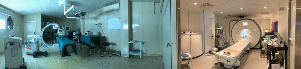
After long years TRUMPF TruSystem 7500 is running with in the neurosuite at
Shmaisani hospital starting from 23-March-2014
LooksCam II Xenosys in the run starting from 14-March-2021 with
SheerVision TTL x4 magnification.
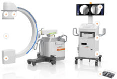
Cios-Spin flat panel in the run.
Notice: Not all operative activities
can be recorded due to lack of time.
Notice: Head injuries and very urgent surgeries are also
escaped from the plan .