Inomed Stockert Neuro N50. A versatile
RF lesion generator and stimulator for
countless applications and many uses
Multigen RF lesion generator .
30-MAY-2002 HAMAD AL-NAEMAT 35 YEARS
WIDE SPREAD AVM OF THE RIGHT OCCIPITAL LOBE WITH FEEDERS FROM THE
RIGHT ACA, MCA AND PCA.
Comments
It was better to treat the patient
conservatively and operate him in better neurologic
condition, but he showed deterioration, for what surgery was
performed as life saving measure.
Despite the fact, that the AVM was huge,
but the foot motor area was the most affected, as noticed in
the follow up.
Anamnesis
The patient was transferred from Grease after
convulsion with loss of consciousness and dense
left hemiplegia to Queen Alia Hospital
23-May-2002.
On examination: The patient has dense left sided
hemiplegia, in Foley's catheter, bedridden with
difficult verbal communication. CT-scan done
03-May-2002 showing bleeding of the right occipito-parietal
lobes, fulfilling the right Sylvian and
perichiasmatic areas. 4-vessel angiography performed
at General Hospital of Athena 16-May-2002, showing the
massive AVM with the feeders from right ACA, MCA
and PCA. MRI with MRA done at Al-Khalidi Medical
Center 25-May-2002 showed massive AVM with
feeders from the right ACA, MCA and PCA with
massive edema reaching anterior to the
sensorimotor strip right side with hematoma with
escalation of the edema in comparison to
previous CT-scan. The
patient was given medications to improve his
condition, but he continued to deteriorate. The
patient then transferred to Shmaisani hospital
for surgical intervention.
Wide right
fronto-parieto-occipital craniotomy with the
flap extending to the left of mid and posterior
third of the SSS and reflected to the right ear.
The dura was widely opened to see all the
pathologic arteries and veins. Dissection of the
pathologic arteries started from tributaries of
the right MCA at the Sylvian fissure. The
pathologic arteries were isolated, coagulated
and bisected. The feeders from the pericallosal
arteries were followed interhemispheric,
coagulated and bisected. It was possible to find
the boundaries of the AVM which was followed and
all feeders coming from posterior circulation
were coagulated and bisected and the
conglomerate of the AVM cluster was removed.
Strict hemostasis and closure. The patient was
sent to the ICU in ventilator but after several
hours started to show conning, for what urgent
CT-scan was performed showing huge extradural
hematoma. The patient was taken another time to
the operating room and the hematoma was removed,
which was from the bone flap and the dura was
opened to evacuate the xanthochromic CSF. The
flap was waxed and the wound closed and the
patient extubated.
Smooth postoperative recovery.
He was sent to the ICU and gradual recovery of
his condition over several days took place.
Follow Up
The patient came to the clinic 13-July-2002 with
left sided spastic hemiplegia. MRI of the brain
done 19-August-2002 showing hydrocephalus
and no evidence for AVM. He progressed myositis
ossificans right hip for what he was advised to
be seen by orthopedics.
The patient then came 06-December-2002 with
slight improvement of the plegia to paresis . He
had convulsions 2 weeks ago.
The patient then came 16-February-2003 after
performing manipulation of the left hip under
G.A, He is convulsion free with continued
improvement of his left sided paresis.
The patient then came 17-March-2009 walking with
crutches with movement all muscles with no
rigidity, except for severe weak dorsi and
planterflexion left foot 0/5. MRI of the brain
with MRA done 18-December-2009 showed the no
evidence of AVM with cavity fulfilling the
previous AVM. That was the last visit of the
patient.
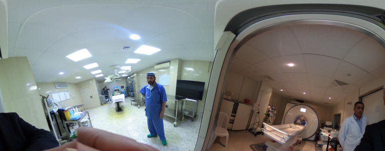
Skyra MRI with all clinical applications in the run since 28-Novemeber-2013.
Inomed Riechert-Mundinger System, with three point
fixation is the most accurate system in the market. The microdrive and
its sensor gives feed back about the localization.
Inomed MER system
Leica HM500
The World's first and the only Head mounted Microscope.
Freedom combined with Outstanding Vision, but very bad video recording and
documentation.
After long years TRUMPF TruSystem 7500 is running with in the neurosuite at
Shmaisani hospital starting from 23-March-2014
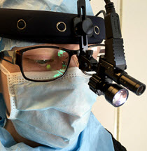
LooksCam II Xenosys in the run starting from 14-March-2021 with
SheerVision TTL x4 magnification.
Notice: Not all operative activities
can be recorded due to lack of time.
Notice: Head injuries and very urgent surgeries are also
escaped from the plan .