Inomed Stockert Neuro N50. A versatile
RF lesion generator and stimulator for
countless applications and many uses
Multigen RF lesion generator .
30-SEPTEMBER-2019 ILHAM AHMAD JURIYE 70 YEARS
AGGRESSIVE RECURRENCE OF LEFT PTERIONAL MENINGIOMA WITH INTRACRANIAL, LEFT
INTRAORBITAL AND RIGHT ETHMOIDAL EXTENSION WITH BLIND LEFT EYE AND EXOPHTHALMUS.
Anamnesis
The patient was operated by me 26-November-2012
for left pterional meningioma with gross
practical radical resection of the tumor. MRI
done 20-March-2013 confirmed total resection of
the tumor with sinusitis of the frontal area
left side. The patient then came
02-December-2013, telling that she still having
hyperlacrimation of the with slight edema of the
lateral part of the superior wall of the orbit.
The patient was sent for investigations and MRI
done 02-December-2013 showing the sinusitis with
a carpet of meningioma at the superior wall of
the left orbit. The patient was advised to
repeat investigations after 3 months. The
patient then came 23-June-2014 with progression
of the exophthalmus left eye and
hyperlacrimation. The left pupil is reactive,
but more wide than the right and she can see
with normal OMNs function. MRI done the same day
showing considerable recurrence of the
meningioma behind the left orbit and she was
advised to undergo surgery for this mass. The
patient disappeared and came came to the clinic
07-August-2019 telling that she is blind in the
left eye for 4 years with pronounced left
exophthalmus and decreased mobility of the eye
movements to all directions. MRI performed
27-June-2019 showing small intradural
compartment over the left frontal area and huge
intraorbital tumor 52x33x25.6 mm pushing the
globe downward with the optic nerve and other
compartments involving the left ethmoidal area.
The patient was sent for cardio evaluation and
new MRI performed 20-august-2019 ruling out
involvement of the carotids.
Bifrontal craniotomy with
reflection of the flap to the right ear. The frontal
sinus was violated to obtain the most lower
projection to the area avoiding by that traction
injury to the brain. The dura
was involved by the tumor at the left side, for what
it was removed. The lateral and superior wall of the
left orbit were dissected and removed in one block.
It was tumorous and it was sent to boiling for 30
min to kill the intraossal tumoral components. A
huge rubbery tumor was seen occupying the
intraorbital superior part. Dissection of the tumor
off the normal tissues with piece-meal resection.
The left anterior clinoid was removed and the tumor
was resected until no apparent tumor masses were
seen. The old lyodura was free of any tumor. The
tumor spread to the ethmoid sinuses were removed. A piece of muscle was
embedded to the frontal sinus . It was impossible to
water-tightly close the dura, for what 2 big pieces
of lyodura were covered over both frontal lobes and
covered by Surgicele. Some places were stitched
using nylon 6 zero. Routine closure of the
wound after stitching the bony elements with ready-Vac drain under the skin flap.
Smooth postoperative recovery. She was sent to the
ICU.
Follow Up
The patient could move the eye to all directions
and could feel the light after resolution of the
ecchymosis.
The patient was discharged the 7th postoperative
day. The histological result was that of
meningothelial meningioma.
Comments
The tumor is increasing in size for what
resection is preferable. She lost vision for 4 years and
recovery of the vision is doubtful. At least for cosmetic
appearance surgery was intimidated.
Skyra MRI with all clinical applications in the run since 28-Novemeber-2013.
Inomed Riechert-Mundinger System, with three point
fixation is the most accurate system in the market. The microdrive and
its sensor gives feed back about the localization.
Inomed MER system
Leica HM500
The World's first and the only Head mounted Microscope.
Freedom combined with Outstanding Vision, but very bad video recording and
documentation.
After long years TRUMPF TruSystem 7500 is running with in the neurosuite at
Shmaisani hospital starting from 23-March-2014
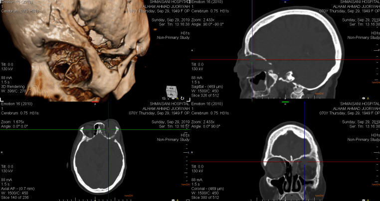
CT-scan with 3D reconstruction with bone defects after the first
surgery.
Coronal MRI showing the involved left
orbit.
Notice: Not all operative activities
can be recorded due to lack of time.
Notice: Head injuries and very urgent surgeries are also
escaped from the plan .