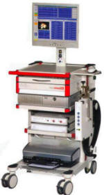
Inomed Stockert Neuro N50. A versatile
RF lesion generator and stimulator for
countless applications and many uses
Multigen RF lesion generator .
11-NOVEMBER-2007 ISSA ABDEL-HAMEED AYOUB AL-HAJ HASAN 56 YEARS GIANT
GLIOBLASTOMA MULTIFORME RIGHT FRONTO-TEMPORO-PARIETAL LOBES.
Anamnesis
The patient came to the clinic 06-November-2007
with headache and neck pain from the right for 1
month with progressive course, with weak left
lower limb. MRI done 05-November-2007 showing
glioblastoma right temporo-parietal lobes. The
MRI was of bad quality.
On examination, the patient is right handed with
left hemihypalgesia and paresis more the distal
muscles both left upper and lower limbs. The
patient was sent for another MRI, which
confirmed the diagnosis and MRA showed the
involvement of the right MCA and its tributaries
inside the mass with massive edema and midline
shift of the brain to the left. The son was
asked separately to gather the family and
detailed discussion about the situation was
performed. They were asked not to hurry with
their decision and to discuss the matter with
all the members of the family. They decided to
let the patient undergo surgical resection of
the tumor with maximal possible resection. The
patient is a known hypertensive in concor 5 mg a
day. He was admitted 10-November-2007 and
operated the next day.
A wide fronto-temporo-parietal craniotomy with
reflection of the bony flap to the right ear was
performed. The dura was stony tight and 100 gm
Mannitol and 80 mg Lasix was administered with
16 mg Decadron. A slight decrease of the dural
tension was noted. The ISIS Inomed highline ion
was used and PRESP was used and epidural mapping
was performed, which showed were the pre and
postcentral sulci are. The dura was opened over
the temporal lobe and partial decompression of
the tumor was was achieved. More relaxation was
noted. While extending the dura incision, the
brain became more edematous and mapping was
performed to see exactly where the central and
postcentral gyri are located. They were pushed
anteriorly.
Temporal lobectomy was performed and the
uppermost part of the tumor was seen with the
MCA branches which were pushed upward and the
tumor through them was removed with preservation
of their continuity. The inferior horn of the
right temporal lobe was violated and seen with
CSF coming from there. The Sylvian cistern was
dissected of the tumor and the branches of the
right MCA were hanging free in the tumor cavity.
The tentorial edge was seen to be occupied by
the tumor and using the arachnoid, the cleavage
was used to remove the tumor parts pushing the
brainstem. Part of the frontal lobe anterior to
the motor area was violated to regain more ample
to the edematous brain, but colleagues and the
general thinking was that performing frontal
lobectomy was not that good option. The MCA and
its branches were irrigated with Papaverine and
the PRESP was repeated and confirmed that the
pre and postcentral gyri still functioning with
the amplitude of the motor area N20 is low as at
the start of the operation, but still present.
Hemostasis with water-tight closure of the dura
and the wound. Ready-Vac drain left under the
skin.
The patient extubated after surgery with deep
left sided hemiplegia, which started to resolve
partially within the next hours.
The patient obeyed commands after 90 min of
extubation and CT-scan was performed 2 hours
later, which showed the tumor cavity with air
and fluid (Saline and hematoma inside the tumor
bed), with hematoma in the frontal area and the
midline shifting is decreased in relation to the
preoperative data.
Follow Up
The patient in next postoperative day was doing
well until he progressed PGE attack. Serial
CT-scan of the brain performed immediately after
surgery and 2 hours before the attack and
immediately after the attack were the same with
residual blood at the bed of the resected tumor.
It is worthy to note, that in these serial
CT-scans the edema of the right occipital lobe
is regaining more intense and wide-spread
character. The patient was given Tegretol over
the previously prescribed Epanutin.
At 10.00 p.m. 12-November-2007, the patient
progressed decerebrating attacks, for what he
was urgently taken to the operating room and the
bony flap was reflected. The dura was stony
tense and the dura was opened first at the
temporal region, through which the lacerated
temporal lobe came out through the small
incision. Another small incision over the most
anterior part of the frontal lobe was performed.
through which the blood clot came out.
Lacerotomy of the temporal lobe and the anterior
part of the right frontal pole was undertaken.
Both incisions were extended to be parallel to
the inferior edge of the bone defect. The clot
above the MCA candelabra was removed with
preservation of the tiny feeders. The previously
mapped cortical areas were in good shape and
appearance and started to give cardio-pulmonary
pulsation and the CSF started to flow from the
posterior horn and the sylvian cistern. Strict
hemostasis with application of Surgicele in the
surgical field. External drain was inserted to
the temporal cavity and other to the frontal
area.
The idea of removing the bone flap was
abandoned, since the brain regained relaxed
appearance. The bone was reflected back to its
original place, after covering the dural
incision by lyodura.
The patient was put in ventilator and the
morning of 13-November-2007 another CT-scan was
performed and the hematomas disappeared and the
shift decreased.
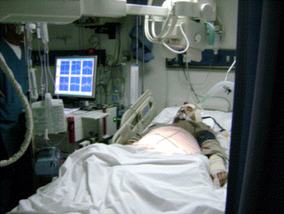
The patient was put in Inomed Highline ISIS
monitor, using ICU-AEP-SEP protocol for 24 hours
and the parameters were stable.
The patient was kept in ventilator until
17-November-2007 and weaning was successful. The
patient showed dense left side hemiparesis. The
patient the next day 18-November-2007 obeying
commands and moving right side of the body and
moving the left upon pain stimulation. The
external drains were removed.
22-November-2007: The patient is clinically
improving and he is still in NGT feeding with
the Chaine-Stokes breathing pattern decreasing
and he is for three days in air room and serial
CT-scan of the brain showed decrease in the
midline shift with appearance of the sulci in
the right parietal region. Slight movement of
the left limbs upon painful stimulation and
communicating well with the surrounding. The
amount of aspirated fluid from the subgalial
area is decreasing. Physiotherapy started three
days ago and he can tolerate setting position
for 2-3 hours twice a day.
25-November-2007: the patient started to
deteriorate with difficult breathing and he was
put in ventilator with dormicum 10 mg/h to
control the epileptic activity and it was
noticeable, that he got sensory aphasia.
The patient dressing showed huge amount of
tumorous fluid coming out under the skin flap
with around 100-200 ml daily.
04-December-2007: the patient still in
ventilator with stable vital signs with the same
neurologic condition and the tumorous collection
still aspirated and waiting for Gliadel to
insert it to tumor bed in hope to stop the rapid
tumor activity. Tracheostomy is planned during
that.
Comments
The patient has the most malignant tumor
of the brain with giant size. Controversy still have place
in what to do exactly and this is governed by several
factors, among them are paramedical ones.
Subtotal resection can help in temporal
resolution of the problem, but the chances for long survival
still remain minimal.
Removal of the insular part of the tumor was
the most difficult and hazardous, because the tumor was highly
vascular and it was difficult to distinguish the right MCA
candelabra from the feeders and SEP was of no help to decide
exactly the degree of the motor function and application of
Papaverine did not help. This is clearly mentioned in chapter 15
of Deletis V. in Neurophysiological Monitoring 2002 edition.
SEP was recorded from both sides and it was
acceptable, despite the fact that, the patient had dense paresis
in the left side of the body.
PRESP can help mapping the brain, but it cannot predict the
outcome of the surgery. MEP is more informative.
For more detailed information about glioblastoma multiforme,
please click
here!
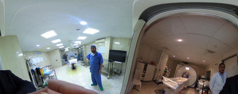
Skyra MRI with all clinical applications in the run since 28-Novemeber-2013.
Inomed Riechert-Mundinger System, with three point
fixation is the most accurate system in the market. The microdrive and
its sensor gives feed back about the localization.
Inomed MER system
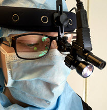
Leica HM500
The World's first and the only Head mounted Microscope.
Freedom combined with Outstanding Vision, but very bad video recording and
documentation.
After long years TRUMPF TruSystem 7500 is running with in the neurosuite at
Shmaisani hospital starting from 23-March-2014
LooksCam II in the run starting from 14-March-2020
Notice: Not all operative activities
can be recorded due to lack of time.
Notice: Head injuries and very urgent surgeries are also
escaped from the plan .