|
The patient came to the clinic 30-January-2006 complaining right
sided weakness with dysarthria sudden onset 18-January-2006. He is
known hypertensive for 7 years in capoten 50 mg three times a day,
tenormin 100 mg once daily modeuretic once daily and baby aspirin
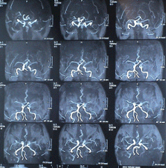
once daily. The patient was sent for MRI investigations and
cardio-consultation. The patient came 01-March-2006 and
stenting of the coronaries were performed including stenting of the
left carotid. The patient neurological status showed slight
deterioration, but the hypertension became milder in severity.
The patient came 26-April-2006 with progressing deterioration
with difficulty in speech and spastic right hand with
difficult walking, nominal aphasia, acalculia, right hemihypalgesia
and hemiparesis more the upper limb.
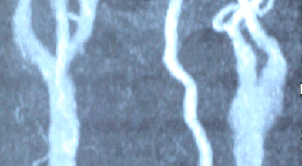
The patient was sent for another investigation. MRA showed
complete occlusion of the left ICA and partial of the proximal
segment of the ECA.
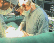
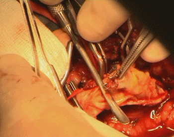
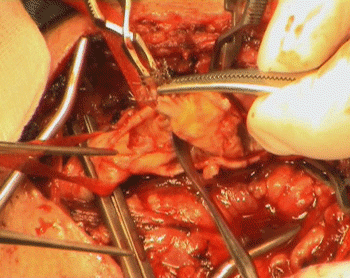
Considering the progressive deterioration of his neurological
course, carotid atherectomy of the left ICA was advised. The patient under G.A with nasal intubation, in case he needs high
dissection, were performed. Incision was made to expose the distal 3
cm of the CCA and the ICA until the upper edge of the stint was
felt. The ECA was dissected and the superior thyroid artery. All was
done with the BP of the patient kept at 170/100 mm Hg. and
continuous cover of the patient with 500 units of heparin/hour.
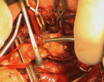
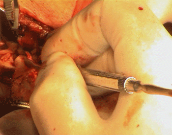
An-Argyle-like tube was prepared in case, but when it was found
that, the back flow of the ICA was weak, it was decided that, no
need for such shunting. For technical details of the operative
details, you can refer to
this article.
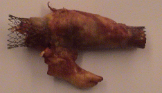
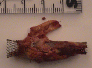
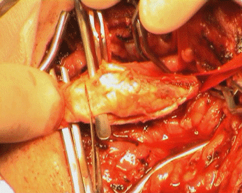
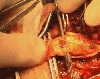
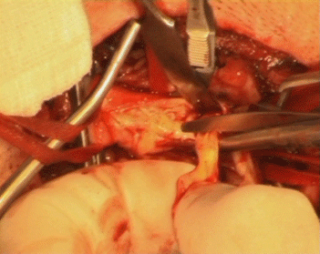
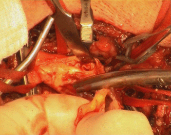
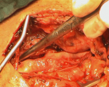
The atheroma was
completely occluding the soft construct of the stint, with minimal
clot inside the very shallow space inside the compressed stint.
After removal of the stint, it regained its cylindrical
configuration, as seen in the lower pictures.
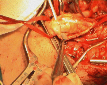
Water-tight closure of the vascular wall with 6 zero nylon and
the carotid bulb and major branches were checked for the flow and
presence of bleeding points. Meticulous heamostasis and ready-vac
drain No 8 left in the wound.
Prompt postoperative recovery, and the patient immediately showed
mild recovery in his speech and the power of his right hand. CT-scan
of the brain was performed immediately after surgery to rule out
progression of hematoma. The patient kept in the ICU for heparin
infusion 650 units/hour and for strict observation of his vital
signs. Comments: Stinting is
a good thing, but it is still needs many corrections in the
technology. As we know the carotid bulb wall has a strong wall
capable of constricting the stint with furthermore atheroma
formation inside the shallow compressed stint as in the sample
before me, which I removed it.
To resolve this problem, my advice is to make the stint from 2 parts
intermingled with each other. The first is what is in the production
now and the second part to be interweaved in the first half of the
construct to offer 2 advantages. The holes will be less wide,
eliminating the progression of the atheroma inside the stint, second
to aid the strength of the construct, to maintain the patency of the
lumen. It seems from the case shown, that the complete occlusion was
the result of these 2 factors.
|