|
The patient came to the clinic 22-February-2006 complaining of right
sciatica for 2 months with positive cough sign down to S1 territory.
MRI done 2 days ago showed extruded disc L5-S1 with right downward
migration with bulge L4-5 disc. On examination, the patient was
limping with SLRS 30 degrees in the right, hypalgesia right L5
territory with weak planter and dorsiflexion right foot. The patient
was advised to undergo surgery, but he escaped.
The patient came 29-April-2006 urging for surgery, because his
pain escalate, that he could not sleep for 5 nights. The patient was
sent for another MRI, which showed the same picture.
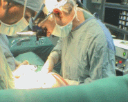
Right L5-S1 hemiflavotomy with S1 root foraminotomy was done and
the extrusion was removed lateral to the axilla. Meticulous cleaning
of the disc space was done from the right side. A tube was inserted
to the disc space and irrigation of the disc space did not gave any
further disc material due to smallness of the hole, which
intentionally was small. The volume of water which was injected to
fill the cavity was 1 ml. It was replaced with gentamicin. Routine
closure of the wound
Comments:
1. This last procedure gave a new idea to decrease the incidence
of recurrence. Neuroendoscope will replace the this tube to perform
multiple or one wide defect in the anterior wall of the annulus
fibrosis under direct vision, to make the inside migrating disc
material more liable to slip anteriorly, decreasing by this way the
recurrence rate. This is a plan for the near future. |