Inomed Stockert Neuro N50. A versatile
RF lesion generator and stimulator for
countless applications and many uses
Multigen RF lesion generator .
23-JANUARY-2022 JIRA NIZAR SULAYMAN 16 YEARS MASS
RIGHT OCCIPITO-TEMPORAL LOBES WITH MASSIVE EDEMA.
Anamnesis
The patient came to the clinic 25-December-2021
complaining of episodes of absences for 6 months
with occurrence once per month with headache.
On examination, the patient complaining of
headache and she was neurologically free. There
is only exaggerated deep reflexes in the right
upper limb.
The patient was sent for investigations and
MRI of the brain done showing a mass in the
right occipito-temporal area, multiple
consistency with massive edema around it,
involving the entire right hemisphere with
deviation of the mid structures to the left with
pending subfalcine herniation. Spectroscopy done
ruling out malignant nature of the mass. It had
pathological arterial feeders from the choroid
plexus right posterior horn. The report was
suggesting mixed solid and cystic lesion with
enhancing solid component suggestive of
pleomorphic xanthoastrocytoma, less likely
intermediate grade astrocytoma.
In concord position with the
right occipital area hanging high, using the
navigation, craniotomy of the right
occipito-temporal area was achieved. The
dura was opened parallel to the upper edge of
the transverse sinus and extended anterior to
lower most of the middle fossa. The tumor was
rubbery solid and the SONOCA 300 could not help
removing the tumor. The tumor was rich in
vascularity and it was needed to coagulate the
tumor and sharp dissection was proceeded. Fresh
frozen biopsy was inconclusive, but it was
advised to remove the whole tumor. The vein of
Labbe was identified and resection of the tumor
was proceeded, so as to preserve this important
vein. The boundaries of the tumor were
identified and the lepto-meningial extension was
considered to be removed with the solid parts of
the tumor. The right posterior horn was seen and
the tumor with the pathologic arteries were
bisected and removed. Radical resection of the
tumor was achieved and confirmed with
intraoperative MRI with T1 mprage with contrast.
Strict hemostasis with preservation of all the
running veins at the tentorium. The brain
regained normal pulsation and relaxed. For more
security, a layer of Surgicele was applied at
the tumor boundaries and over the prominent
veins at the tentorium. Routine closure of the wound. Smooth
postoperative recovery. She was sent to the
ICU for 24 hour observation.
FOLLOW UP
Too early now, but the patient in the ICU 6
hours later alert and no neurologic deficit.
The final histologic result was
intracerebral schwannoma with no evidence of
malignancy. This case to the mentioned reference
is the 12 reported case in the literature.
Comments
The patient has strange mass with such
massive edema. Surgical removal was mandatory.
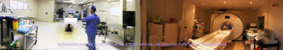
Skyra MRI with all clinical applications in the run since 28-Novemeber-2013.
Inomed Riechert-Mundinger System, with three point
fixation is the most accurate system in the market. The microdrive and
its sensor gives feed back about the localization.
Inomed MER system
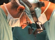
Leica HM500
The World's first and the only Head mounted Microscope.
Freedom combined with Outstanding Vision, but very bad video recording and
documentation.
After long years TRUMPF TruSystem 7500 is running with in the neurosuite at
Shmaisani hospital starting from 23-March-2014
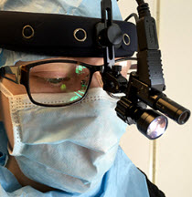
LooksCam II Xenosys in the run starting from 14-March-2021 with
SheerVision TTL x4 magnification.
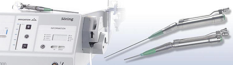
SONOCA 300
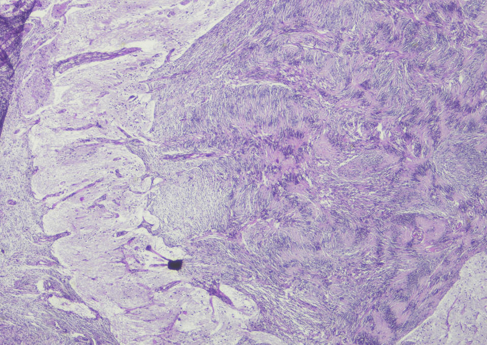
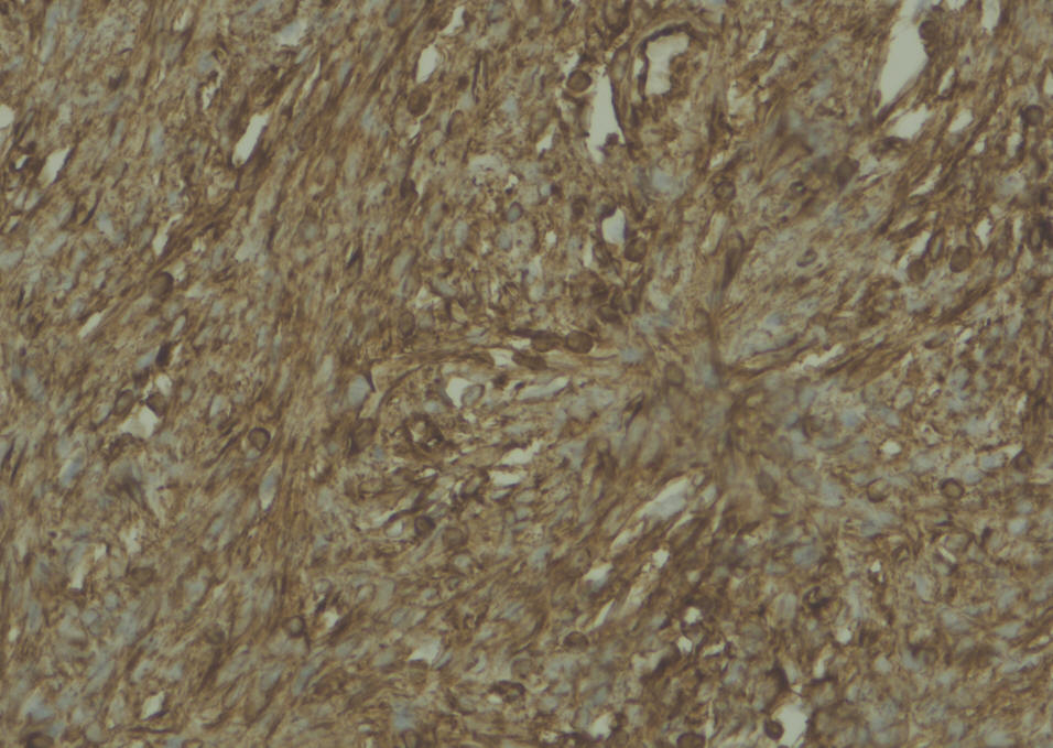
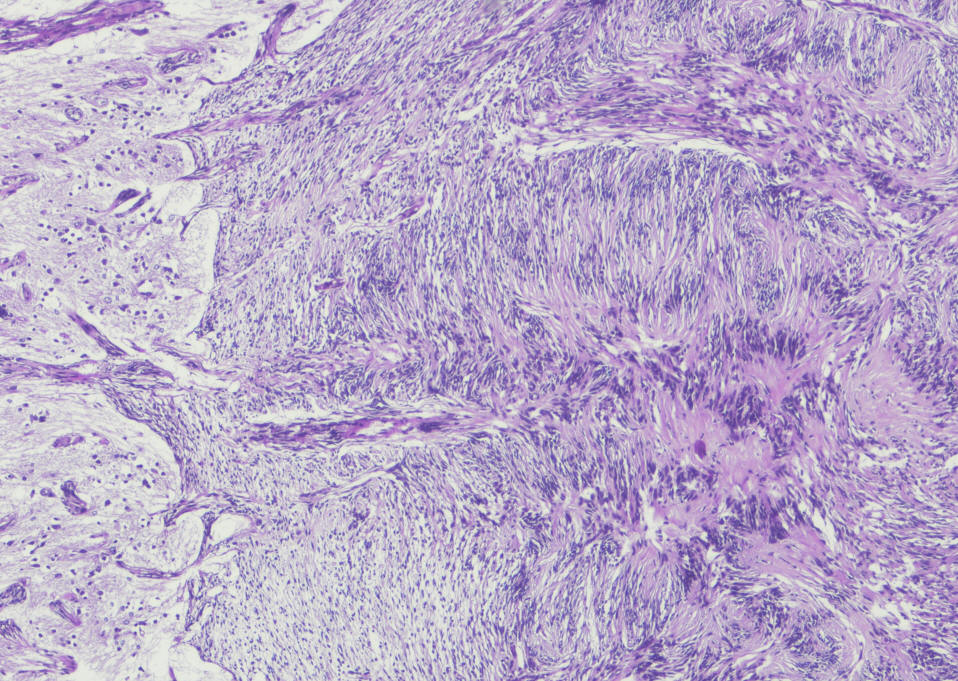
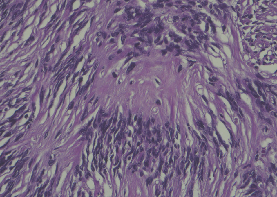
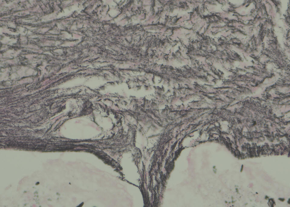
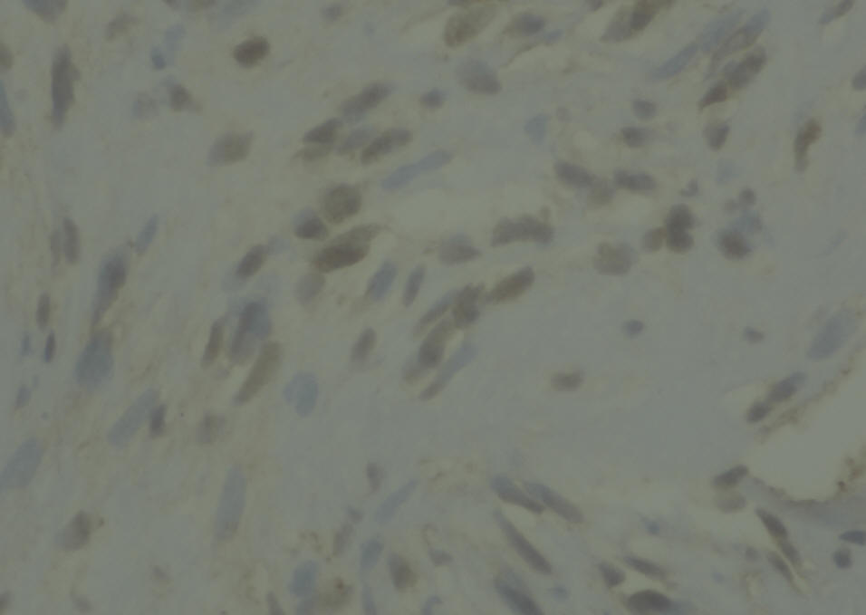
Sections show a circumscribed tumor composed of spindle cells
displaying prominent nuclear palisading and Vercoy bodies. This
lesion exhibits peripheral tongues extending into the brain cortical
tissue. There is no evidence of mitotic activity, nuclear anaplasia
or necrosis. Tumor shows heavy interstitial reticulin deposition and
reacted positively to S-100 and Vimentin. Tumor did not react to
GFAP and EMA. Proliferative index was estimated at 2%, but not
exceed 3% in any area. There is no evidence of malignancy.
Conclusion: Intracerbral schwannoma. ( Prof. Yahya F. Dajani
Consultant pathologist. 29-January-2022.
Notice: Not all operative activities
can be recorded due to lack of time.
Notice: Head injuries and very urgent surgeries are also
escaped from the plan .