www.neurosurgery.tv 
   
Munir Elias 20-12-2013

 
Most of the site will reflect the ongoing surgical activity of Prof. Munir Elias MD., PhD. with brief slides and weekly activity. For reference to the academic and theoretical part, you are welcome to visit  neurosurgery.tv


The patient  36 years age got gun shot 18 months ago with subsequent paraplegia  and anaesthesia below D9. There was complete absence of spinal cord function below this level, which was confirmed by ECS and EMG. He underwent recently operation for major bed sore 1 month ago and huge rotational skin flap was rotated from the right posterior aspect of the leg. He pass urine with regular voiding with condom and regular defecation without laxatives.

A detailed discussion about the kind of surgery and the pros and cons and the limitations of such procedure were explained to the patient and he was told that despite the final result, which could be achieved within 3-5 years, he may be in need for orthoses to make him able to walk. The patient agreed in these terms and he was operated.

Skeletonization of D7 down to the sacrum was performed. The transverse processii of L1-2-3-4 were exposed and the intertransverse ligaments were removed. The lower edge of the transverse ligaments were drilled, including the isthmus and upper part of the lateral masses from both sides to expose the lateral edge of the whitish ligamentum flavum edge, which must be partially removed to expose the running nerve root and the ganglion and postganglionic part

The operation took 20 hours and the patient received 4 units of packed cells and FFP and kept in the ICU for 6 hours. The patient was covered by targocid 400 mg daily and pain-killers. The ready-vac drain was removed after 24 hours and dressing of the spine was performed for 4 days, till it became clean. He was able to set on the wheelchair in the 5th postoperative day.

20-January-2006: The patient and wife noticed that all the mass reflexes, which caused problem to the patient before the operation disappeared. This is mostly due to isolation of the preganglionic part of the lumbar nerves. No deterioration in his bowel and bladder functions were noted.

In my opinion, this fact making the operation giving advantage, even in the early postoperative period, manifesting the disappearance of the negative phenomena of the destroyed spinal cord.

Follow Up:

1. The patient came 26-July-2006 to the clinic. On examination, the patient still with spastic pattern of both lower limbs with condom. There is still no signs of motor recovery, but the crude sensation step downward to the level of the umbilicus. The surprising thing is that the sacrificed dorsal roots all returned their function. ( 10% of each root left in continuity during the procedure, so as to prevent slippage of the roots and to ease the insertion of the anastamosis, as used with the partial resection of the hypoglossal nerve in cross anastamosis for the facial nerve.

2. The patient was told to press his thoracic cage to stimulate the lower limb movement and continue physiotherapy and given medications to be reevaluated after 6 months.

3. Cross-anastamosis in paraplegia below D9 started to give results. The patient operated 1 year ago from Israel came  27-December-2006 to the clinic. ECS and EMG performed showed that there is starting innervation of Th 11 and 12. The patient lower limbs muscles became bulky and he could contract the lower abdominal muscles and some movements in the pelvic girdle. Crude sensation descended down to the inguinal level both sides.


Notice:

The saphenous nerve as seen in the picture is accompanying the femoral artery. The sartorius muscle is a good landmark. It is preferable to localize the nerve at the midthird of the leg and working medial to the sartorius in the upper third up to the point, where all the other motor branches unite. In the lower third the dissection is done lateral to the sartorius muscle  with subsequent opening of the channel, until the nerve start to give branching, usually three subdivisions. The length of the graft is usually 33-35 cm in adult patient


Comments:

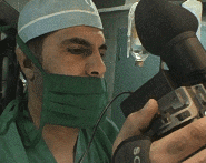
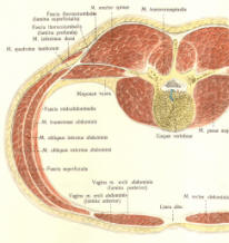
1. As seen in the picture the midline incision is the less traumatic approach to the postganglionic part of the roots. Dissection to go around the paraspinal muscles is not wise and cause more trauma to the soft tissues.

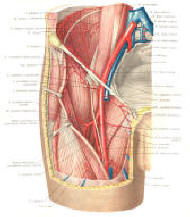
2. The integral part of the operation is not to cause harm to the patient. For that only functionally unimportant neural grafts must be harvested and they are at maximum 4 nerves from each leg: the sural, saphenous, anterior cutaneous branch of the femoral nerve and posterior cutaneous branch of the sciatic nerve, which is missing in this case due to previous operation for the bed sore.

3. Using microscopic facility the grafts were anastamosed using 6 zero nylon. During that the postganglionic truncks were partially resected to leave the posterior wall of the epineurium so as to use it as traction to the anastamostic  site. The number of stitches was governed by achieving perfect alignment of the anastamosis. It was necessary to put between 6 to 10 stitches at each site to regain the goal.

4. Routine closure of the wound and as seen the grafts are hanging lax and free above the laminaes with luxury length, to avoid traction in case the patient bend his back. 

5. From the previous part it became evident, that only three major branches could be accessible to be used for grafting for the intended purpose. The posterior cutaneous branch of the sciatic nerve in this condition was impossible to harvest, because plastic grafting of the thighs were performed for the bed sores.

6. This means that such surgery could be applied to paraplegics below D10 to make the bridges to L1-S1  roots. Of course, the paraplegics below D9 can have benefit from such surgery, but with less benefit.

Comments:

1. this is the third performed operation, and with the increased number of the operations  and with time and end results will be clear. The first performed operation was in 28-January-2004 which gave partial but excellent results concerning the anastamosed nerves. He could show the improvement of some muscles and sensation of some roots, but the operation was not organized enough to make him able to walk, due to several factors, among them the negligence of the patient for his situation and disappearance of the patient mostly due to financial reasons. He came only once to me 18 months after the surgery and I was astonished with the good reinnervation of the grafted nerves.

2. The surgical standards are becoming more standardized and the steps of the operation becoming more precise. The maximal 8 grafts harvested govern the limitations of the operation and the number of the lost grafts in the patient also play a major impact in the decision-making.

3. This operation can be applied not only to paraplegics, but also to stationary post-transverse myelitis and other conditions, where the certain roots for good lost their function.

4. The fact that the dorsal roots supply relatively small segments of dermo-myotoms make some skepticism about the final result of the operation and the presence of 2 stitching points to fill the gap and the sensory nature of the grafted neural material, all play  a negative theoretical role in the outcome. Time will tell.


Back Up!


View Larger Map


© [2005] [CNS CLINIC - NEUROSURGERY - JORDAN]. All rights reserved