Most of the site will reflect the ongoing surgical activity of Prof. Munir Elias MD., PhD. with brief slides and weekly activity. For reference to the academic and theoretical part, you are welcome to visit
neurosurgery.tv
Inomed Stockert Neuro N50. A versatile
RF lesion generator and stimulator for
countless applications and many uses
Multigen RF lesion generator .
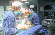
17-OCTOBER-2012 MUHAMED ALI ABU-SBETAN 72 YEARS
SEVERE CERVICAL CANAL STENOSIS C2-3, 3-4 AND C5-6.
Anamnesis
The patient came to the clinic 27-June-2012
complaining of difficult walking for 5 months
with LBP and bilateral sciatica. MRI lumbar
spine performed 06-June-2012 showing bulge
L5-S1. The patient is walking with help of 2
persons. On examination the patient had full
power of the upper limbs and Hoffmann sign left
side. SLRS was 60 degrees in the left with pain
with weak all muscles of the lower limbs 3/4
left leg and right foot and 4/5 of the right
quadriceps muscle with hypalgesia of the left
leg extending 20 cm above the left knee. The
patient then sent for whole spine MRI, which was
done 03-July-2012 showing bulge L3-4 with mild
degree of L5-S1 spondylolisthesis. The old lower
screw still slipped as before after the
performed by me operation
13-April-2012
for huge extruded disc C5-6. The patient did not
perform MRI of the cervical spine and when he
came 26-July-2012 telling that his condition is
dramatically deteriorating with heaviness of the
left upper limb the last 4 days with swelling of
both legs. The patent was resent to complete the
investigations with cardio consultation. The
patient came 29-August-2012 with MRI of the
cervical spine done 22-August-2012 showing
severe cervical canal stenosis of C3-4, C4-5 and
C5-6 with malacia of the spinal cord. The
patient was resent for MRI of the brain and
cardio consultation. MRI of the brain done first
time 09-September-2012 of bad quality and he was
advised to repeat it. It was done
25-September-2012 showing atrophic changes
compatible with age and scattered lacunar
infarctions, more around the left lateral
ventricle. Cardio consultation gave permission
only 13-October-2012 to undergo surgery under
G.A.
On examination: the patient in addition to more
deterioration of previous condition got weak
grip extension and left triceps muscle left
upper limb -4/5. The patient was examined
immediately before surgery and it was clear that
he cannot walk for 5 months and has severe
tetraparesis more pronounced in the left upper
limb and drop right foot.
In supine position with the
head slightly flexed and under traction with 6
Kg, the lamina of C2,3,4,5 and 6 were
skeletonized until the groove of the lateral
masses was seen. Using high speed drill the
laminae were drilled until the most lateral part
abutting the groove of the laminae were seen and
transparent. The drilling was done to include
the lower third of C2 and upper third of C6. All
these structures were reflected off the position
and to the left to avoid any iatrogenic trauma
to the spinal cord and removed in one piece. The
epidural fat was missing and the bridging veins
between the ligamentum flavum and the dura were
coagulated and sharply bisected.
Routine closure of the wound. Smooth
postoperative recovery with normalization of the
power of the both upper limbs and considerable
improvement of the power of both lower limbs.
Please! wait for 3-5 min till the
video start to load. It depends upon the internet
connection.
Comments
The patient has
severe stenosis of the spinal cord starting from
C2-3 down to C5-6. The patient is deteriorating
and only surgical decompression was the only
solution to halt the deterioration.
Using drilling and thinning of the lateral parts
of the laminae, give guaranty to avoid
mechanical trauma to the spinal cord during
surgery.
Leica HM500
The World's first and the only Headmounted Microscope.
Freedom combined with Outstanding Vision.
Notice: Not all operative activities
can be recorded due to lack of time.
Notice: Head injuries and very urgent surgeries are also
escaped from the plan .