|
The patient came to the
clinic 04-December-2005 complaining of LBP and
right sciatica with clinical manifestations of
cauda equina syndrome after performed surgery
for PLD L4-5 22-September-2005 elsewhere. |
|
|
MRI of the lumbar spine
performed 15-September-2005 before surgery
showed extruded disc L4-5 right side. MRI
performed 30-November-2005 showing recurrence of
the disc with bigger extrusion. He was operated
3 years ago for the same disc at the same level
and side. |
|
|
On examination at that time: the patient
was limping with scoliotic stance. SLRS was 20
degrees in the right and 30 degrees in the left with pain. There
was weak
dorsi and planterflexion right foot 3/5. |
|
|
The patient was advised to
undergo another surgery and he came
21-June-2008, after performing three further
surgeries for the same recurrence by another two
neurosurgeons elsewhere. |
|
|
MRI of the lumbar spine
performed 10-February-2008, showing still having
recurrence at the same level with total
deformity of the right L4-5 facet joint. |
|
|
On examination: the patient
still in agonizing pain with scoliotic stance
with SLRS 30 degrees in the right and 40 degrees
in the left with pain. He had weak dorsi and
planterflexion both feet more the right.
|
|
|
The patient was sent for new
MRI with CT-scan and dynamic X-ray studies of
the lumbar spine. |
|
|
The patient then came
24-June-2008 with MRI confirming the recurrence
and practically absent right L4-5 facet joint. |
|
|
The patient was advised to
undergo surgery for the recurrence and to remove
the flail fragments of the totally destroyed
facet and to use MTF allograft to accelerate the
fusion between L4-5. |
|
|
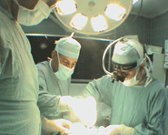
Right L5 foraminotomy with
bilateral flavotomy of the L4-5 level was
achieved under visual control of
image-intensifier. In the right side of the
dural sac, there was a massive scar embedded
with movable bone fragments, which were the
remnants of the right L4-5 facet. All these
structures were removed and the dura was
inspected to be fee from these pain-generating
elements. |
|
|
The extruded disc which was
rubbery hard was removed and drilling of the
disc space of L4-5 was done to reach the
anterior part of the soft tissue material, which
was removed subsequently. |
|
|
Through this tunnel, chips of
MTF bone allograft
were pushed to fill the intradiscal cavity. They
were inserted with impactor to have good
resistance and to prevent backward slipping. |
|
|
The right L4 root and the L5
roots were exposed to visually eliminate all
compressive elements. |
|
|
Routine closure of the wound
and smooth postoperative recovery with
normalization of the power of both feet. |