|

Most of the site will reflect the ongoing surgical activity of Prof. Munir Elias MD., PhD. with brief slides and weekly activity. For reference to the academic and theoretical part, you are welcome to visit
neurosurgery.tv
Functional Neurosurgery
functionalneuro.surgery
Functionalneurosurgery.net
IOM Sites
iomonitoring.org
operativemonitoring.com
Neurosurgical Sites
neurosurgery.art
neurosurgery.me
neurosurgery.mx
skullbase.surgery
Neurosurgical Encyclopedia
neurosurgicalencyclopedia.org
Neurooncological Sites
acousticschwannoma.com
craniopharyngiomas.com
ependymomas.com
gliomas.info
gliomas.uk
meningiomas.org
neurooncology.me
pinealomas.com
pituitaryadenomas.com
Neuroanatomical Sites
humanneuroanatomy.com
microneuroanatomy.com
Neuroanesthesia Sites
neuro-anesthessia.org
Neurobiological Sites
humanneurobiology.com
Neurohistopathological
neurorhistopathology.com
Neuro ICU Site
neuroicu.info
Neuroophthalmological
neuroophthalmology.org
Neurophysiological Sites
humanneurophysiology.com
Neuroradiological Sites
neuroradiology.today
NeuroSience Sites
neuro.science
Neurovascular Sites
vascularneurosurgery.com
Personal Sites
cns.clinic
Spine Surgery Sites
spine.surgery
spondylolisthesis.info
paraplegia.today
Stem Cell Therapy Site
neurostemcell.com

Inomed Stockert Neuro N50. A versatile
RF lesion generator and stimulator for
countless applications and many uses

Multigen RF lesion generator .
|
 |
|
 |
| |
|
Introduction
Neurodegenerative diseases include a very wide group of
disorders affecting the central nervous system (CNS).
Many of these disorders arise from the combined effects
of genetic predisposition and environmental factors.
This results in reduced cognition (e.g. Alzheimer’s
disease, dementia with Lewy bodies, and vascular
dementia), motor system performance (e.g. amyotrophic
lateral sclerosis), or both (e.g. Parkinson’s disease
and prion diseases). In general, neurodegenerative
diseases show a wide diversity of etiology and a broad
clinical phenotype spectrum, but all have in common the
decrease in neuronal function and neuronal cell death
due to activation of apoptotic pathways, programmed cell
death, or other mechanisms. |
|
The evolution of magnetic resonance (MR) techniques for
imaging the CNS has led to significant advances in the
understanding of brain changes associated with
neurodegenerative disorders. Conventional MR imaging
(MRI) provides detailed anatomic information with
excellent tissue contrast and spatial resolution.
Additional MR sequences can provide information
concerning tissue metabolism, water diffusion, and
perfusion. All together, these MR modalities have been
demonstrated to be relevant in the clinical evaluation
of neurodegenerative disorders for early diagnosis,
differential diagnosis, and monitoring of disease
activity. MR spectroscopy (MRS), in particular, has
provided insights into some of the metabolic
abnormalities associated with neurodegeneration, thereby
helping to elucidate the underlying pathophysiology of
these disorders, although it has yet to have significant
impact in routine clinical usage. The goal here is to
describe the common findings of MR spectroscopy (MRS) in
this field and its limitations. We will focus on proton
MRS (1H-MRS), as the majority of MRS studies in
neurodegenerative disorders utilize this technique and
it can readily be performed as part of a routine MR
study. It should be stressed, however, that other nuclei
(such as 31P or 13C) have been used in research studies
of various neurodegenerative disorders.
Dementia
Dementia is a clinical diagnosis defined as a decline in
memory and other cognitive functions that affect the
daily life in an alert patient. Major causes of dementia
include Alzheimer’s disease (AD) and vascular dementia
(VD) and, less commonly, frontotemporal dementia and
dementia with Lewy bodies. Consensus clinical criteria
have been applied for diagnosis of different dementias,
but the sensitivity and specificity of these criteria
are variable. The diagnosis of dementing disorders by
means of the current clinical criteria remains difficult
and, occasionally, due to the mixed pathology existing
in the brain of demented patients, the underlying cause
of dementia cannot be definitely determined even after
the histopathologic examination of the brain.
The difficulty in making diagnoses exclusively based on
the current clinical criteria has generated the
incentive for identifying specific neuroimaging markers
for various dementing pathologies. Thus, in vivo changes
of demented brains have been studied with different MR
modalities with increasing frequency.
In this context, 1H-MRS has been shown to be useful in
the differential diagnosis of dementing illnesses, as
well as in monitoring early disease progression and
effectiveness of therapies.
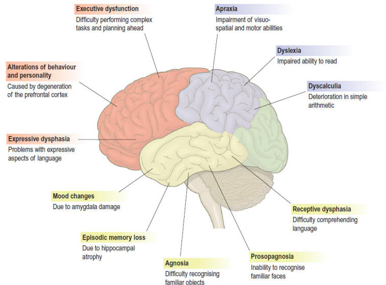
Fig.1 Clinical features of dementia. This figure shows
some of the common features of dementia that are caused
by degenerative changes in different parts of the
cerebral cortex (frontal lobe = red, parietal lobe =
blue, temporal lobe = yellow). The clinical picture in a
particular person depends upon the distribution and
severity of the changes. From Paul Jones-Clinical
Neuroscience. |
|
|
 Skyra MRI with all clinical applications in the run since 28-Novemeber-2013.

Leica HM500
The World's first and the only Headmounted Microscope.
Freedom combined with Outstanding Vision, but very bad video recording and
documentation.

After long years TRUMPF TruSystem 7500 is running with in the neurosuite at
Shmaisani hospital starting from 23-March-2014 |