Most of the site will reflect the ongoing surgical activity of Prof. Munir Elias MD., PhD. with brief slides and weekly activity. For reference to the academic and theoretical part, you are welcome to visit
neurosurgery.tv
Inomed Stockert Neuro N50. A versatile
RF lesion generator and stimulator for
countless applications and many uses
Multigen RF lesion generator .
24-NOVEMBER-2010 IGHZAYEL RAFDAN AL-DOSARY 30
YEARS HUGE SUPRA RETRO AND PARASELLAR MASS WITH INVASION OF THE III
VENTRICLE, AFTER FAILED ATTEMPT OF TRANSSPHENOIDAL REMOVAL 06-AUGUST-2010
ELSEWHERE WITH HISTOLOGIC RESULT OF CRANIOPHARYNGIOMA.
Anamnesis
The patient
came to the clinic 20-November-2010 complaining
of loss of vision left eye for three weeks with
decreased vision right eye. She is complaining
of headache and amenorrhea for 8 months.
The patient was operated in Saudi Arabia
06-August-2010 with any benefit. Transsphenoidal
approach was used and biopsy revealed
craniopharyngioma as the histological result.
On
examination: The patient is almost blind
in left eye and decreased vision right eye with
hemianopia temporal field. The patient has no
sensory, nor motor deficit. She is amenorrheic
and feel thirst all the time.
MRI brain done 13-November-2010 showing huge
tumor with intra-supra- parasellar extension
with invasion of the III ventricle. It was clear
to see the previously performed approach and to
see the small bone defect at the pituitary
floor. The patient was sent for
neuroophthalmological and endocrine evaluation
which confirmed the almost blind left eye and
the the residual of the visual field of the
right eye. The cortisol ACTH where very low.
Prolactin was 51 ng/dL.
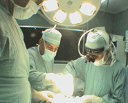
Bifrontal approach was achieved with reflection
of the bone flap to the right. The dura was
opened parallel to the inferior edge of the bone
defect. The olfactory bulbs and tracts were
mobilized from the mediobasal frontal lobes. The
right olfactory tract was dissected down to the
trigone. The tumor was attacked between the
optic nerves and piece-meal resection was
performed. The tumor was rubbery in consistency,
but it had good cleavage. That part compressing
the left optic nerve was removed totally. The
posterior right parasellar extension was removed
in second stage. The suprasellar tumor which was
invading the III ventricle was removed totally,
trying to avoid traction with force. The
pituitary stalk was the origin of the
craniopharyngioma, which was attacked at the
last stage, trying to minimize the remaining
dust at the pituitary stalk, without violating
its anatomical integrity.
Routine
closure of the wound. Smooth postoperative
recovery and improvement of the vision in both
eyes. The patient was happy with her
postoperative improvement.
Conclusions:
This case and similar 4 cases
over 30 years of experience with solid component
of the craniopharyngioma pushing up far the III
ventricle with blindness in one or both eyes,
means that tumor is not only distorting the
optic nerves, chiasm, A1 segments and the floor
of the III ventricle, it is compressing them
with force. The one stage total removal in
these cases must be avoided so as not to end
with such catastrophe. The flow of surgery
in this case was smooth and the solid part came
with ease from the floor of the III ventricle.
In the next case I will divide such surgery for
2 stages: The first will be the removal of the
intrasellar, antesellar and parasellar parts and
to leave the suprasellar part intentionally, to
avoid such catastrophe. After that, with in 1-2
months later according to MRI data, to see the
downward migration of the suprasellar part over
the days by the cardio-pulmonary pulsation to
the empty cavity created by the first surgery.
If the tumor is separable from the floor of the
III ventricle, then to proceed to the second
stage of removal. If it is stuck with the floor,
then better to leave it and think about
radiotherapy.
Minirin in some patients is
not effective, and there is a notice, that the
previous trans-sphenoidal approach could
participate with this no response.
With the experience with DI,
Minirin is effective and the DI usually is
responding well to medication. This hidden DI
which showed itself one month ago and now, did
not reflect itself by the specific gravity of
the urine. The specific gravity was 1.010 before
surgery and 1.005 during the peak of
hypernatremia.
The hypothalamus is still a
mystery and there must be unknown to us
functions, that need to clarify and special
management for them.
The cause of DI and
hypernatremia, by no means could be explained by
the reaction of the pituitary stalk. It is due
to reversal of the floor of the III ventricle
and the infendibulum down to its normal position
over 3-4 hours of decompression and resection of
the solid part of the tumor.
Please! wait for 3-5 min till the
video start to load. It depends upon the internet
connection.
Comments
The patient underwent first
transsphenoidal surgery for craniopharyngioma,
which was originating from the pituitary stalk
and expanding to the III ventricle and
compressing the chiasm and both optic nerves and
the right ICA. This mission is impossible, even
with advent of the most robust technologies.
With bifrontal approach with my own
modification, patented 1985, it is possible to have these
findings and make it possible to practically remove the
whole tumor, preserving during that, the involved pituitary
stalk and the olfactory function.
Follow
Up
The patient the morning of
25-November-2010 showed somnolence and status
epilepticus, for what, CT-scan urgently was
performed, which revealed no hematoma and no
residual of the tumor. The patient urine output
was more than 6 liters during night with Na
275mmole/L. Blood sugar was over 400 and the
patient was urgently put in ventilator to
control the status epilepticus with propofol and
depakine I/V was started and the Epanutin was
stopped and Tegretol was started in NGT. Minirin
was started but she showed minimal response to
Minirin. Trying over the next hours to
control the dangerous level of hypernatremia was
impossible despite all precautions to slow the
slope of regression. It became 178mmole/L
the next day.
The morning of 27-November-2010 showed
clinically nearly brain stem areflexia, for what aggressive
measures where taken to decrease the brain edema. Another
CT-scan was performed and the patient showed return of her
corneal, gag reflexes and she could breath spontaneously in
the SIMV mode. With all efforts to avoid rapid regression of
the hypernatremia, it became 145 during this day. She showed
improvement of brain stem functions the night of this day.
The morning of
28-November-2010 the patient showed clinical
picture of brain stem areflexia and the BP
started to decrease, which was corrected with
dopamine infusion. Propofol was stopped and all
antiepileptic drugs. Inomed ISIS IOM was
connected to the patient over 24 hours, which
confirmed the absence of cortical activity and
profound decrease in AEP and VEP, but with good
response to SEP and MEP. Clinical brain death
was established in mid day of 29-November-2010.
The patient still in the ventilator.
Notice: Not all operative activities
can be recorded due to lack of time.
Notice: Head injuries and very urgent surgeries are also
escaped from the plan .