|
 During 25 years, several hundreds of repeated operations for recurrent lumbar disc prolapse were performed by me, and they were escaped the records. During the last five years, I started to pay special attention to the causes and predisposing factors for the recurrence of the first and second time. My experience cannot reflect the actual incidence, since most of the repeat surgeries, were performed by other neurosurgeons, but the recurrence rate of the surgeries performed by me were
around 10%, and the recurrence after the second operation, was also
10%. A fifth recurrence was noted in 2
cases, since, as usual, the second recurrence take place after complete collapse of the disc space with subsequential fusion of the two adjacent bodies several months later,
but it happens that, this not the case
and the recurrence was noted to be
present even after the fourth surgery. During 25 years, several hundreds of repeated operations for recurrent lumbar disc prolapse were performed by me, and they were escaped the records. During the last five years, I started to pay special attention to the causes and predisposing factors for the recurrence of the first and second time. My experience cannot reflect the actual incidence, since most of the repeat surgeries, were performed by other neurosurgeons, but the recurrence rate of the surgeries performed by me were
around 10%, and the recurrence after the second operation, was also
10%. A fifth recurrence was noted in 2
cases, since, as usual, the second recurrence take place after complete collapse of the disc space with subsequential fusion of the two adjacent bodies several months later,
but it happens that, this not the case
and the recurrence was noted to be
present even after the fourth surgery.
During the last 4-5 years it was clear
that the recurrence depend heavily upon
two factors, that is the height of the
disc space from where the extrusion took
place and the dimensions of the defect
of the annulus fibrosis from where the
extrusion happened. If the disc space is
still high and the defect of the annulus
fibrosis is wide, then the expected
recurrence must be more than 20% of
estimation.
Simeone F.A., mentioned that, each surgeon should keep a careful record of recurrences. It is to be hoped that the surgeon will maintain good enough clinical relations that patients will feel free to return if their symptoms develop again. When patients are told before the first operation that a recurrence could develop at any time, they seem to tolerate better the news that
recurrence took place but they will be
kept under constrain thinking about it.
It became a common policy, to send the patient to perform check MRI with contrast, if his complains persist more than month and the neurological deficit is not regressing. It happened, even in the third day and the next week after the operation. Most of them were negative for recurrence, but, when the patient knows, about that, his psychological and neurological state start to improve. Some cases, with MRI confirmed absence of recurrence, and in the case that they, continue to suffer, another check MRI in some cases, showing the recurrence. It mean that intradiscal mass displacement was the cause of pain, and, at last, the recurrence became evident.
Trying to understand the causes of the recurrence , to avoid second surgeries, many variations of surgical techniques were performed, and they all failed to resolve the problem. Noticing that, most of the recurrences after other neurosurgeons, usually by the absence of scar and the natural history of the patient after the first surgery, it is clear in some cases, that missing the extrusion, usually the migrating parts, were the cause for second surgery. This group was the easiest, since you practically operating the patient as be the first operation.
Trail to maximal cleaning of the disc space with removal of wide part of the posterior part of the annulus fibrosis, did not slow down the recurrence rate. In contrast, after several operations, I got the feeling that, this approach exposes the patient for more chance to the intradiscal mass displacement to take place to the direction of the wide annular defect.
Trail to maximal cleaning of the disc space through very small holes, usually, that one, through which the first extrusion took place, using very small diameter pituitary rongours, with trail to coagulate the defect to minimize the defect, also did not help, since the posterior part of the annulus, became part of the recurrence.
Trail to insert big piece of bone in the disc space, configured, so as not to slip back, sometimes was rewarding, but I have cases with the graft slipping back with the annulus fibrosis with subsequent agonizing pain and evolution of permanent neurological deficit.
The dilemma is running around an objection, that for such "minor problem" , is to go for cages or fusion devices. Even with those, the outcome is inferior to simply repeat removal of the recurrence, and it happened that these cages slipped instead of the annulus fibrosis. This indicate that the epiphysis is playing a hidden role in these tragic scenarios.
The actual incidence of recurrence is highly variable. In one of the longest follow-up series, involving 984 patients with a mean follow-up period of 10.8 years, the recurrence rate was 6 percent, and one-third of these recurrences developed during the first year after operation. When, studied specifically, the recurrence rate has varied from 3 percent to 19 percent, generally being higher in series with longer follow-up times.
The management of recurrent disc herniation remains somewhat controversial. Many surgeons believe that fusion should be considered at the time of second operation, although this concept is held more widely among orthopedic surgeons than neurosurgeons. I do not at present believe that recurrent disc herniation is an indication for fusion. As experience with better imaging techniques evolves, and as individual surgeons develop confidence in their procedure, the idea that a fusion must be done to relieve symptoms becomes less relevant. In the past, when the mechanics of the spine and the role of lumbar spine surgery were less clearly understood, fusion was a way to immobilize a segment where repeated difficulties evolved. Now that better imaging techniques are available, the specific cause of the symptoms can be discovered, and when it is indeed a recurrent disc herniation, removal of the disc fragment should be enough to relieve symptoms. Herron treated 46 patients for recurrent disc herniation at an average of 7 years after the previous laminectomy. In this group, 28 patients (69 percent) had a good result, 10 (24 percent) had a fair result, and 3 (7 percent) had a poor result. Fusion was not used in any case. Herron concluded that in the absence of objective spinal instability, recurrent disc herniation can be treated adequately by repeat discectomy.
Starting from August-2009 after long
time of hesitation, PEEK Satellite
nucleus replacement sphere was adopted
in all personal lumbar disc surgeries.
The technique is convincing, that it
will decrease the recurrence of sphere
complication down to 2%. This will
resolve the recurrence in young aged
patients , but patient younger than 17
years and old patients with osteoporosis
still not candidates for such surgery.
PEEK Satellite nucleus replacement
sphere not only decreases the recurrence
of the disc, but also keep the disc
space height in its original status and
prevent stress in the corresponding
facets and give some movement to the
involved segment.
CASE PRESENTATION
Case-1
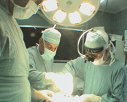
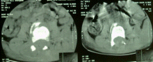
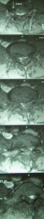
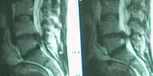
A gentle married lady 45 years came 28-03-2004 complaining of LBP for 9 years. She got exacerbation of pain the last 2 months with left sciatica, down to L5 territory with positive cough sign. On examination she had weak dorsiflexion toes both feet, more in the left. The patient was sent to MRI lumbar spine, which showed the L4-5 extrusion. The patient was operated 17-04-2004. Left hemiflavotomy with foraminotomy for left L5 root was performed and removal of the extrusion was done with left sided cleaning of the disc material was undertaken. The patient was seen several times after surgery with normalization of the feet power and SLRS was 90 degrees both sides. The patient, then came 13-06-2004 complaining of sudden attack of left sciatica for one week with numbness of the heel of the left foot. The patient was neurologically free during examination, but for assurance, she was sent for check MRI with contrast
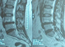
 MRI done 21-June-2004, as seen in the inferior row, showed a massive recurrence, even with intradural migration of the annulus fibrosis. Due to relatively minor complains of the patient, she agreed with hesitation for the second surgery, which was undertaken 04-07-2004. During surgery, some points could be considered: 1.Usually we transfer the epidural fat to the maximally involved
areas by compression, aided with transferring the subcutaneous fat in pedicle to the epidural space. This fact made dissection for the second surgery more easy and the fibrous tissue was fatty in appearance. 2. The extruded material was a layer from the annulus fibrosis. It was removed very gently and the tail, which was intradural came the last in one piece, after what the CSF start to flow from the dural defect , which was about 2 mm in diameter and stitched accordingly. 3. At the end of operation, another subcutaneous fat was transferred with pedicle to the epidural
space . Video recording available. The patient was discharged the second postoperative day without complications. Case-2:
 |
 |
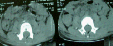
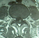
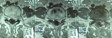
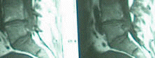
A young lady 37 years age was seen 29-06-2004 , complaining of right sciatica for two weeks .She was operated elsewhere 1999 for PLD L4-5 for left sciatica. The patient was brought in wheelchair with agonizing pain with SLRS 10 degrees in the right and 45 degrees in the left with drop right foot and weak dorsiflexion left foot and planterflexion both feet. She has hypalgesia below the right knee and dripping urine for two days. MRI lumbar spine with contrast done 24-06-2004 demonstrating huge recurrence at the same level. The patient was operated 30-06-2004: Right hemiflavotomy with drilling of the upper 2/3 of the right L5 lamina was done with right L5 root foraminotomy was done. The extruded annulus fibrosis was removed in one piece. Cleaning of the disc space from the right was performed and good amount of disc material was removed . The left side of the operative field was not violated , since there was dense scar, and fat transfer with pedicle was performed to the right side. |
 |
 |
Postoperative period was uneventful, and the patient was discharged walking the second postoperative day with complete normalization of the neurological state. |
Case-3:
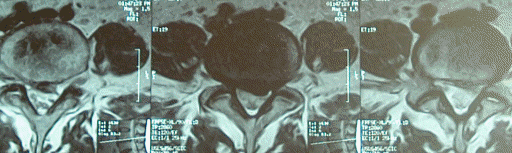
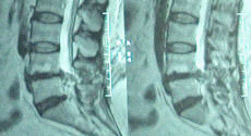
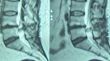
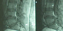
57 years age patient came 07-07-2004 complaining of left hip pain with with numbness of the left foot for two weeks, after setting for three days in a chair for long time during mourning ceremony . The patient was operated for PLD L4-5 1985 by me for left sciatica. He had recurrence 5 years later and was operated by another neurosurgeon 1990 for the same sciatica. Then, he was seen by me 18-09-2003 and MRI with contrast performed 17-06-2003 showing a huge recurrence at the same level with left downward migration of the annulus fibrosis (see fig.)
The page was revised 16-November-2009.
|