Inomed Stockert Neuro N50. A versatile
RF lesion generator and stimulator for
countless applications and many uses
Multigen RF lesion generator .
31-JANUARY-2007 SAWSAN ISMAEEL AL-TAMIMI 43 YEARS WIDE-SPREAD RIGHT
FORNTO-TEMPORO-PARIETAL INTRAOSSAL MENINGIOMA.
Anamnesis
The patient came to the clinic 16-January-2007
complaining of a bony lump in the right
fronto-parieto-temporal region progressing in
size. She noticed this several months without
pain. She brought with her brain CT-scan with
the diagnosis "fibrous dysplasia". Considering
her age, unusual localization of the process for
dysplasia, which usually invade the base of the
skull, and rapid growth rate, intraossal
meningioma was suspected and MRI was requested.
MRI with contrast performed, showing typical
intraossal meningioma with wide=spread invasion
of the dura with carpet covering the three lobes
with epicenter 3 cm above the right pterion.
A wide question mark incision was
made and the bone flap was created 2 cm away from
the pathologic bony structure. The center of the
abnormal bone was bleeding profusely, and bleeding
was controlled by bone wax.
The bone flap was elevated and sent for thermal
deproteinization with temperature 125 for 15 min.
The pathologic dura was removed with the meningioma
carpet, which was abutting the anterior edge of the
bone defect.
After thermal coagulation of the bone flap, it was
more clear, that the pathologic bone was extending
anteriorly, for what secondary bone flap was created
anteriorly to make sure the radical resection of the
tumor.
The suspected dura was also removed and 4 pieces of
large size lyodura were necessary to use to close
the dural defect.
The boiled bone was remolded by the high-speed drill
so as to regain a normal-looking appearance. The
hyperostotic bone was marble-like in consistency.
The 2 bone flaps were fixed together by nylon, and
glue. The flaps were put back in place and fixed.
and routine closure of the wound.
Smooth postoperative recovery.
She was sent to the ward.
Comments
Fibrous dysplasia usually start early in
life with young males and it invade the base of the skull.
The dura under the fibrous dysplasia is normal and has no
pathologic components.
Intraossal meningioma has dural involvement, with a carpet,
which could be more wide than the hyperostotic part of the
bone.
To prevent recurrence, it is mandatory, to remove all the
involved dura and take if feasible 1-2 cm away the
pathologic margins of invasion, as in this case.
It is unnecessary to use bone cement or metallic mesh, as in
this case, the bone was deproteinized with thermal exposure
of the bone flap for 15 min in the autoclave with
temperature 125 degrees, Celsius.
During the 27 year period more than 20 operations were
performed, returning the boiled bone flap to place. An
interesting phenomenon, is the disappearance of the bone
flap in the usual skull X-rays in the first 6 months, then
its reappearance, despite the fact, that the flap is holding
normal in place. It could be explained by the gradual
wash-out of the calcium and then the gradual filling of the
mineral after this period.
For further information about meningiomas, please click
here!
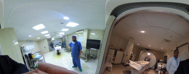
Skyra MRI with all clinical applications in the run since 28-Novemeber-2013.
Inomed Riechert-Mundinger System, with three point
fixation is the most accurate system in the market. The microdrive and
its sensor gives feed back about the localization.
Inomed MER system
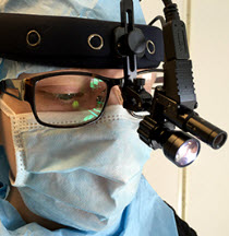
Leica HM500
The World's first and the only Head mounted Microscope.
Freedom combined with Outstanding Vision, but very bad video recording and
documentation.
After long years TRUMPF TruSystem 7500 is running with in the neurosuite at
Shmaisani hospital starting from 23-March-2014
LooksCam II in the run starting from 14-March-2020
Notice: Not all operative activities
can be recorded due to lack of time.
Notice: Head injuries and very urgent surgeries are also
escaped from the plan .