|
The patient came to the clinic 07-March-2006 with flail right upper
limb. The patient is an Iraqi citizen got RTA 02-August-2004 with
avulsion injury to the right upper limb and fracture of the right
humerus mid-third. The patient underwent several operations for the
fracture and neurolysis.
On examination, the deltoid , supra-infraspinatus , biceps
brachii muscles are completely paralyzed. The median and ulnar
nerves are completely not functioning. The triceps is weak 4/5 and
the brachioradialis muscle is 2/5.
MRI performed 07-March-2006 showing avulsion of right C7 root and
ECS confirmed extensive injury of the right brachial plexus with
absent response from right C6-7 and T1 roots.
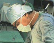
The patient underwent exploration of the
brachial plexus
through the most ugly incision undertaken before. The pectoralis
major was bisected for 3 cm to have more superior access to the
brachial plexus. The median nerve was identified from the most
distal part and followed up to end with vigorous scan without neural
elements.
The brachial artery and vein were dissected and preserved, trying
during that to preserve the integrity of the lymphatic nodes and
channels.
The ulnar nerve was identified distally and followed proximally.
It was penetrating the bone with useless neural elements at the
lower third of the arm. Considering these findings, that the median and ulnar nerves are
completely destroyed at the distal segments, they were cut were
acceptable fibers were noted.
The MCN was exposed and studied. It
was weakly transmitting stimulation to the long head of the biceps
muscle, for what it was left in place intact.
The deltoid muscle was acceptable during direct stimulation of
the muscle. Considering that, the radial nerve was preoperatively
functioning, no attempt was made to explore the radial and axillary
nerves.
The skin incision was extended inferiorly in the chest area to
expose the intercostal nerves: Th2, 3, 4 and 5. Considering the low
level of injury of both median and ulnar nerves, it was needed to
harvest both sural nerves 35 cm length both. The were divided in 2
parts to have four bridges.
Cross anastamosis was performed between Th2 and 3 to the median
nerve and Th4 and 5 to the ulnar nerve. The suturing was performed
under microscopic facility.
Routine closure of the wounds. Ready-vac drain left in the chest
wound for 24 hours. |