Inomed Stockert Neuro N50. A versatile
RF lesion generator and stimulator for
countless applications and many uses
Multigen RF lesion generator .
06-AUGUST-2020 WALEED MUHAMED SALEH AL-SAADI 36
YEARS NEUROFIBROMATOSIS I WITH HUGE NEUROFIBROMA ORIGINATING FROM LEFT C3
WITH EXTRAMEDULLARY AND HUGE PARAVERTEBRAL EXTENSION.
Anamnesis
The patient came to the clinic 26-July-2020
complaining of limitation of neck movement for
all directions due to huge mass in the left
posterior aspect of the neck a the
craniovertebral junction. He noticed it 6 months
ago and it is growing by time. MRI done
22-July-2020 showing a huge left paravertbral
mass with intradural compartment at the level of
C3 shifting the spinal cord to the right.
On examination: The patient has limitation of
neck movement to all directions except flexion
of the neck. The mass is firm unmovable,
residing under the base of the skull posterior
to the mastoid. Inspection of area for presence
of abnormal veins, which could be a clue for
malignancy is absent. The patient was
neurologically free.
The patient was sent for investigations and MRI
with MRA and MRV of the area was done confirming
the presence of neurofibroma, originating from
the left C3 root, pushing the spinal cord to the
right, expanding the left C2-3 foramen, pushing
the left vertebral artery anterior and upward.
ICA and jugular veins are normal. The huge
extravertebral part is multicompartment with
several consistency, reaching the C5 level.
Midline incision with laminectomy
C2,3 more to the left. The dura was opened above the
tumor and radical removal of the intradural part of
the tumor with involved roots was achieved. The left
extraforaminal huge part of the tumor was of
multiple consistency with one huge stony consistency
rounded mass were removed. The tumor was severely
vascularized, that it needs 5 units packed cells and
10 units FFP during surgery. All the visible parts
of the tumor were removed. Routine closure of the
wound with Ready-Vac drain inside the wound.
Smooth postoperative recovery.
He was sent to the ICU since the operation took
around 10 hours with massive intraoperative
bleeding.
Follow Up
The final histologic result was that of
malignant peripheral nerve sheath tumor. The
tumor cells show, in the better differentiated
area, focal reaction to S 100. Proliferative
index by Ki 67 shows hot areas with increase to
levels exceeding 15% especially in perivascular
zones and less differentiated component. Tumor
cells were non-reactive to Sox 10 (positive in
only 2/3 of such tumors). The report was
received 27-August-2020. The report was issued
by Prof. Yahia F. Dajani.
The patient came 10 days after surgery. He was
neurologically free with normal healing of the
wound.
The report was given upon family request
27-August-2020 to be re-evaluated by Hussein
Cancer Center for possible radio, chemotherapy
or both.
Comments
The patient is in need for surgery to
prevent spinal cord damage and to make the patient able to
move his head.
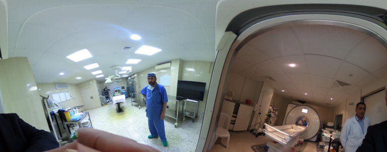
Skyra MRI with all clinical applications in the run since 28-Novemeber-2013.
Inomed Riechert-Mundinger System, with three point
fixation is the most accurate system in the market. The microdrive and
its sensor gives feed back about the localization.
Inomed MER system
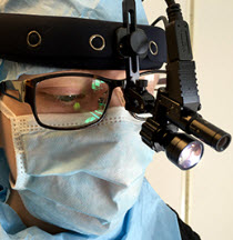
Leica HM500
The World's first and the only Head mounted Microscope.
Freedom combined with Outstanding Vision, but very bad video recording and
documentation.
After long years TRUMPF TruSystem 7500 is running with in the neurosuite at
Shmaisani hospital starting from 23-March-2014
LooksCam II in the run starting from 14-March-2020
Notice: Not all operative activities
can be recorded due to lack of time.
Notice: Head injuries and very urgent surgeries are also
escaped from the plan .