|
The patient came
to clinic 11-November-2006 complaining of headache for 3 years
with disturbed memory. The condition deteriorated and she got neck
pain and vomiting attacks for 2 weeks and she came with dexametasone
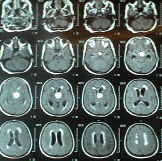
coverage. CT-scan performed 05-Noveber-2006 showed a
suprachiasmatic mass in the left lateral and III ventricle with
secondary acute hydrocephalus. On examination: the patient had
left upper limb weakness and hypalgesia. There was mild weak right
lower limb.
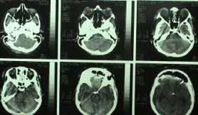
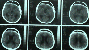
The patient was sent for MRI of the brain with contrast and brain
MRA and MRI of the cervical spine. MRI performed the next day
showing a huge mass resembling craniopharyngioma. The mass was
mainly cystic with extraventricular extension left to the third
ventricle. It was pushing the hypothalamus to the medial side. It
had left para and retrosellar extension.
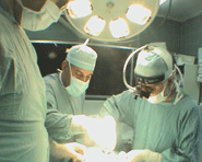
The patient was operated: bifrontal subfrontal with modification
to be combined with left pterional approach was achieved.
Complete mobilization of the left olfactory tract down to the trigon
and partial mobilization of the contralateral olfactory tract was
performed, after what the frontal lobes felt down by gravity, making
it easier to explore the chiasmatic region.
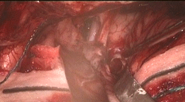
After dissecting the arachnoid around the left olfactory trigon,
the prechiasmatic cistern and the left carotid cistern were sharply
opened. It was possible to see the solid part of the
craniopharyngioma pushing up the left ICA and stuck with it,
resembling a giant aneurysm. Further retrochiasmatic dissection
carried out and medial to the left olfactory trigon, the A1 was
running tightly pushed anteriorly. Posterior and above the
left A1 the cyst was attacked and puncture of the cyst wall was
performed to rule out presence of giant aneurysm. A golden greenish
fluid came out. It became clear that the lesion is a
craniopharyngioma. Evacuation of the huge cyst was achieved. A small
miniretrator was inserted directly to the cavity and all the debris
and calcified elements were remove. The cavity was
extraventricular, pushing the III ventricle medially. There was no
CSF flow from there. The most posterior part of the calcifications
was stuck with the basilar artery. A small remnant left there in its
wall to avoid possible spasm.
The left parasellar part was debulked and a small remnant was
left adherent to the lateral wall of the ICA. The oculomotor nerve
was dissected sharply from the tumor and the calcification was
removed including the antero-lateral wall of the carotid cistern,
which was involved by the tumor.
Ommaya reservoir was put subgalial and its proximal tip inserted
to the evacuated cavity. Inspection of the subchismatic region
showing the pituitary stalk hanging free and had no relation to the
pathology. Routine closure of the wound and smooth postoperative
recovery.
The patient showed some dilatation of the left pupil which
normalized after 4 hours. Despite the anatomical preservation of
olfaction the patient was checked for olfaction. She could not
differentiate odors.
16-November-2006: the patient is ambulating with normalization of
the oculomotor nerve function and she can feel and differentiate the
odors in both nostrils. There were not diabetes incipidus, nor
psychomotor irritation. She was transferred to the ward from the
ICU.
Comments:
1. Despite the practical subtotal resection of the
craniopharyngioma, it is preferable to leave Ommaya reservoir
inside the tumor bed to make it easy for the patient to evacuate the
fluid in case of possible recurrence.
2. The olfactory function with anatomical preservation of their
integrity, could loose function temporarily, but they then gradually
regain function.
3. Wide exposure in this case made it possible to attack the lesion
from all corners. The anterior lower edge of the bony defect was
flush with the base, avoiding any traction injury to the midiobasal
structures and yielding a good visualization space.
4. The left olfactory trigon was stuck to the chiasm, despite that
it was possible to do surgery medial and lateral to these
structures.
5. You can refer to the theoretical data about craniopharyngioma,
click here please
for that. |