|
The patient came 22-August-2006 complaining of severe exophthalmus
left eye with blind left eye and light sensation on the right eye.
The patient was operated 4 times for meningioma elsewhere 08-July-2003,
17-November-2003, 08-April-2004 and 15-April-2006. Loss of vision
left eye for three years. Deterioration of vision right eye the last
20 days. MRI performed 15-August-2006 showed wide spread intraossal
pterional meningioma with invasion of the tuberculum sellae and left
retrobulbar extension and ethmoidal growth with involvement of both
cavernous sinuses and reaching the tenorium both sides.
On examination: the patient is practically blind, but with some
light perception on the right eye. All oculomotor nerves were
disturbed both sides with dilated both pupils. Olfaction was
preserved. Hearing loss left ear and right facial central
paresis with right hemiparesis.
Considering that the vision on the right eye is rapidly
deteriorating with the presence of chiasmal compression and
unacceptable exophthalmus, it was decided to operate him. The
patient was admitted 3 days prior to surgery and anemia and
hypoalbuminemea were corrected and diabetes incipidus was noted and
corrected.
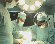
Bifrontal craniotomy was performed, using the old incision,
which was extended by using the old bony flap and the nasion with
part of the anterior part of the left orbital roof were skeletonized
and the involved by the tumor were exposed to high temperature
in autoclave for 10 min to kill the tumor cells. The left
tumorous huge pterion was drilled completely, exposing during that
the neurovascular bundle of the left superior orbital fissure. All
the lateral wall of the left orbit was drilled off and drilling was
extended down until the infratemporal fossa was seen. All the
accessible bony elements in that area were drilled off, including
the posterior half of the orbit and the lesser wing. This part was
extradural.
The dura was opened parallel to the anterior edge of the bony
defect at the level of the crista Galli and the falx was bisected.
The left olfactory bulb was scarified and the other was preserved.
The dura in the planum sphenoidale was sharply dissected and
drilling of the planum and tuberculum sellae was achieved, removing
during that the ethmoidal extension of the tumor. During this part
massive arterial bleeding came from the left superior ethmoidal
artery, which was controlled in several stages of removal.
That part of the tumor originating from the tuberculum sellae was
removed, after what it was possible to see the right optic nerve
hanging free and the supraclinoid ICA freely running underneath.
From this point, the left side of the chiasm was identified and the
scarous tumor was removed, leaving small remnants stuck to it and
the adhere to it the A1 segment.
After removal of the left part of the tumor prechasmatically,
it was possible to see the left optic nerve, The postero-medial part
of the orbital roof was drilled off to remove the retroorbital
extension of the tumor.
Inspection of the right lesser wing for tumor presence was
negative. The arachnoid separating the pituitary gland was kept
intact. The extradural left optic nerve was seen pushed downward and
the periorbita was incised down to the annulus of Zinn. Part of the
periorbita was involved with tumor , which was resected.
A huge muscle graft was harvested from the left quadriceps
muscle. Several pieces were inserted snuggly to fill the cavity at
the resected part of the ethmoidal sinus. Another parts were
inserted to fill all the spaces created after drilling the very huge
left pterion.
The bone flaps were gathered and fixed by several means to
reconstruct the frontal part of the face and anterior half of the
left orbital roof. Routine closure. The operation took 12 hours
duration. Smooth postoperative recovery.
27-August-2006: The patient can smell odors and the vision of the
right eye the same, but to my surprise and out of expectation, he
regained light perception of the left eye. I had a similar case with
complete blindness for 9 years in one eye , which regained function
later. This happened 15 years ago and I remember the name of the
patient, but there were no video-documentations to prove that.
Comments:
1. One can ask: why to do such major surgery in this case? The
answer is to improve the vision in the right eye and resolve the
unacceptable exophthalmus. Time will give the answer.
2. For more theoretical data concerning meningiomas please visit
meningiomas.org, or
meningiomas.org. |