|
OPERATIVE VIDEO
GALLERY
TO SEE YOUR OPERATION
CLICK HERE!
Functional Neurosurgery
Functionalneuro,surgery
IOM Sites
iomonitoring.org
operativemonitoring.com
Neurosurgical Sites
neurosurgery.me
neurosurgery.mx
skullbase.surgery
Neurosurgical Encyclopedia
neurosurgicalencyclopedia.org
Neurooncological Sites
acousticschwannoma.com
craniopharyngiomas.com
ependymomas.com
gliomas.info
gliomas.uk
meningiomas.org
onconeurosurgery.com
pinealomas.com
pituitaryadenomas.com
Neuroanatomical Sites
humanneuroanatomy.com
microneuroanatomy.com
Neuroanesthesia Sites
neuroanesthesia.info
Neuroendocrinologiacl Site
humanneuroendocrinology.com
Neurobiological Sites
humanneurobiology.com
Neurohistopathological
neurohistopathology.com
Neuro ICU Site
neuroicu.info
Neuroophthalmological
neuroophthalmology.org
Neurophysiological Sites
humanneurophysiology.com
Neuroradiological Sites
neuroradiology.today
NeuroSience Sites
neuro.science
Neurovascular Sites
vascularneurosurgery.com
Personal Sites
cns.clinic
Spine Surgery Sites
spine.surgery
spinesurgeries.org
spondylolisthesis.info
paraplegia.today
Stem Cell Therapy Site
neurostemcell.com
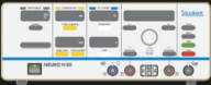
Inomed Stockert Neuro N50. A versatile RF
lesion generator and stimulator for countless applications and many
uses.

MultiGen RF lesion generator
About
Bipolar Pulsed Mode RadioFrequency Applications. Please Click here!

To see first authority,
Click here!
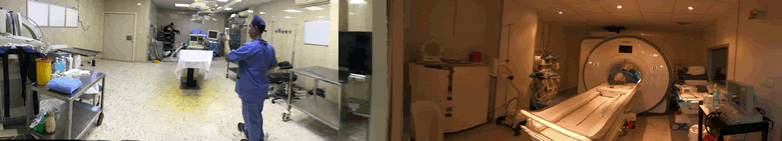
INTRAOPERATIVE MRI RESULTS DURING THE PERIOD
2013-2018 IN NEUROSURGERY AND SPINE SURGERY.
Site Map.
MODIFIED POSTERIOR FOSSA APPROACH
INTRACRANIAL
VASCULAR MALFORMATIONS
|
NEUROSURGERY
This site is directed mainly to the medical
audience and neurosurgeons, partially aimed to present the operative and
academic activities of Prof. Munir A. Elias Shawash over 40 years period. Here
the neurosurgeon can find the standards and new modifications in the treatment
strategies in paraplegia, brain tumor, spinal cord injuries, head injury, pain
management strategies, including neuralgia of different etiologies, movement
disorders. Neurosurgeon needs a very long way to understand that, experience is
important in this field of medicine - neurosurgery.
Stroke and ruptured arterial aneurysms remain in the upper list of difficult
problems, which are far from perfection and the mortality rate remains high.
Spinal surgery is extensive and take 80% of the neurosurgical activities:
prolapsed disc , lumbar, cervical and dorsal are the top ranking in practice
followed by other degenerative spine problems, such as spondylolisthesis, OPLL
with cervical and lumbar canal stenosis.
Neurosurgery has time-sensitive decision-making strategies. This is governed by
the rapidly changing status of the patient. Neurosurgeon must react accordingly
to the recent moment. He must be able to predict, or at least to keep in mind
the possible complications, and react with caution to prevent them, before they
escalate. Intraoperative video documentation made it possible to analyze and
retrospectively discover some causes of complications, which the neurosurgeon
previously blamed himself for that. It became clear, that some triggering
factors for complications are presenting before their eminence.
Here come the power of intraoperative monitoring using IOM ISIS HighLine 32
channel with all available parameters, which can alarm the functional shifts
before they become real disaster and to take the appropriate measures before
they become irreversible.
Neuroanesthesia is the cornerstone in proper navigation of such monitoring to
make it feasible and to guide the patient with safe margins until he pass the
surgical storm. It starts from the preoperative period until the patient is no
more complaining, whatsoever it needs time.
Intraoperative morphological navigation using BrainLab skyvision with the MRI
with the most high standards available with all softwares more empower the
surgeon to know and see all the data and take the proper action and know at
which stage he is standing.
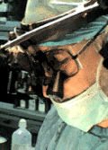
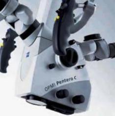
Pentero-C is not only a microscope, it give the neurosurgeon the power to see,
what he could not see before with the appropriate softwares.
Concerning stemcell therapy, it got excellent results in all disciplines, where
tissue have good regenerative potential with primitive biological function. In
central nervous system, which consist of neurons and supportive tissues, the
glial tissues can regenerate, but their role in final higher functions is
somehow limited. Treatment of paraplegia and stokes are still far from
perfection. The tried surgical treatment of paraplegia with putting anastamoses
between the upper dorsal functioning roots and lower lumbar non-functioning
roots gave bad results. It seems resolving such problems must have other
dimensions or combination of them.
Not all new standards in neurosurgery can stand time. Only the good for the
patient's outcome will stand and remain even, if they are too old.
Since 2007 at Shmaisani hospital functional neurophysiologic navigation ISIS
Inomed Highline 32 channels and BrainLab Suite integrated with Siemens Skyra 3
tesla fMRI with fibertraking (DTI) and other more than 80 Syngo softwares for
intraoperative monitoring are in practice since 2013.
It is very sad to say that, very huge medical corporations can misinform the
neurosurgeon about the new products without telling that these items having
disadvantages, but in the contrary, reporting that no morbidity or complications
can arise, until the neurosurgeon discover them in his personal experience.
Profit-oriented corporations must respect the ethics and tell the true story
about any product, so as, at least to be ready to inform the patient and to try
to resolve these possible complications. For that reason, the author started to
be expert in SolidWorks and Autodesk Inventor to create new designs and
studying their efficacy.
When you have one complication, you forget the hundreds of successful
alike surgeries and a sad feeling will overwhelm you, not mentioning
that when you live in the third world, where no body understand this,
including the primitive wrong directed legal system.
Using MATLAB and signal processing and Inomed MER and RM stereotactic
device with micro-macro electrode recordings, we could translate up to
now more than 20 areas of the deep nuclei and surrounding areas of the
brain. This is only the start of the project, which with time must cover
more than billion of sites in the brain and later the spinal cord,
creating the GPS inside the brain and neural structures. This and and
the chemical map of neurotransmitters could jump the humanity to new
level of understanding the normal and pathologic conditions from
different conventional aspects and can lead even to catch the activity
of the functioning brain. It is simple as creating a new language and to
put the letters, and then the poems and literature according to this new
language.
With the introduction of new technologies, new dimensions arise and new problems
also. When you have more data, you have more information to deal with and your
tactics and options also may expand further, but despite that, complications
will remain and they will need solutions. At last we are human beings and the
more effort you do, the more spirit comfort you will feel when your life come to
end.
|
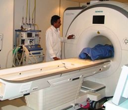
Skyra MRI with all clinical applications in the run since 28-Novemeber-2013.
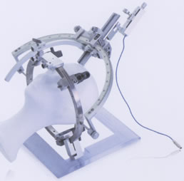
Inomed Riechert-Mundinger System, with
three point fixation is the most accurate system in the market. The
microdrive and its sensor gives feed back about the localization.
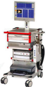
Inomed ISIS IOM
highline with 32 channel and Neuroexplorer version 4.2 is functioning for
several months, starting from 01-August-2007. For more detailed information
about this functional neuronavigation machine with its early alarming signals,
please refer to
inomed.com
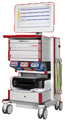
Neurostimulation with ISIS Inomed system.
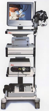
Leica HM500
The World's first and the only Headmounted Microscope.
Freedom combined with Outstanding Vision, but very bad documentation.
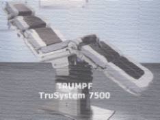
After long years TRUMPF TruSystem 7500 is running with in the neurosuite at
Shmaisani hospital starting from 23-March-2014
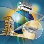
Prestige LP Cervical Disc system Medtronic.


|