Most of the site will reflect the ongoing surgical activity of Prof. Munir Elias MD., PhD. with brief slides and weekly activity. For reference to the academic and theoretical part, you are welcome to visit
neurosurgery.tv
19-AUGUST-2006 KHALED IBRAHEEM MUHAMED
33 YEARS RECURRENT PLD L5-S1 LEFT SIDE.
The patient came 16-August-2006 complaining of LBP for 8 months
with left sciatica. MRI done which is missing and according to that
, he was operated 02-April-2006 elsewhere for PLD L5-S1, after what
the patient claiming that his condition deteriorated and he was in
bed for 21 days with progression of deep venous thrombosis "DVT"
of the left lower limb 2 weeks after the operation.
MRI performed
17-July-2006 showing the "recurrence " of L5-S1 with left downward
migration. On examination: the patient has still DVT of the left
lower limb with agonizing left sciatica, hypalgesia of left L5 and
S1 roots territories. He had weak dorsi and planterflexion of the
left foot.
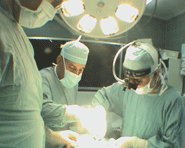
The patient was covered with clexane 40 mg /day before surgery to
prevent the escalation of DVT and was operated. Using the old
scar, which was very low, it was impossible to performed the
operation, for what the skin incision was extended high.
Skeletoniztion of L5 lamina and the upper edge of the sacrum, which
was full of adhesions. The left side of the L5 lamina was
drilled partially to expose the normal dural sheet. Foraminotomy of
the left S1 root was performed to expose the normal looking neural
structures. The bleeding due to clexane made the dissection very
difficult. It was necessary to expose S1 and S2 to be sure about the
normal anatomy. The medial part of the left L5-S1 facet was drilled
to gain direct access parallel to the scarous running root. The
extruded disc material was stuck to the dura and intermingled with
the scar. Piece-meal resection of the extrusion and the scar was
performed from the healthy looking parts until the root became free
of the scar and the extrusion. Meticulous cleaning of the disc space
from the left side lateral to the axilla. Inspection under the
axilla was negative. Inspection of the S1 and S2 roots at their
foramina ruled out any compression. Routine closure. Prompt
postoperative recovery.
Comments:
1. Using clexane
24 hours before surgery with the dose 40 mg made the operation, very
difficult. Even the scar was diffusely oozing, mandating continuous
bipolar coagulation and heamostasis with irrigation of saline. Most
of the time was spent to identify the neural structures and to
continue working with confidence, so as to avoid damage to the
already damaged neural structures.