Most of the site will reflect the ongoing surgical activity of Prof. Munir Elias MD., PhD. with brief slides and weekly activity. For reference to the academic and theoretical part, you are welcome to visit
neurosurgery.tv
Inomed Stockert Neuro N50. A versatile
RF lesion generator and stimulator for
countless applications and many uses
Multigen RF lesion generator .
22-MAY-2008 NAHLAH NABEEL BAKEER 25 YEARS
GIANT CYSTIC CRANIOPHARYNGIOMA WITH PAN-PARA-SUPRASELLAR EXTENSION MORE TO THE
RIGHT.
Anamnesis:
The patient was seen by me
the morning of 22-May-2008. She was admitted to
Al-Shmaisani hospital under supervision of other
neurosurgeon.
The clinical course of the
patient was protracted over 18 months, with
ataxic gait visual disturbances.
MRI performed 5 days ago
showed a giant cystic mass in the sellar region
pushing the floor of the third ventricle ,
causing dramatic ventricular dilatation and
pushing the brainstem posteriorly with the
basilar artery and spreading down over the
clivus.
On examination, the
patient is aphonic and cannot protrude her
tongue with motor aphasia. She was almost
quadriplegic with slight movement of her left
hand. She had hiccup, but the breathing pattern
was acceptable.
During 5 hours of observation
with 9 time reevaluation, the patient was
deteriorating with conning pending. The
breathing pattern start to deteriorate, for what
she was taken urgently to the operating theater.
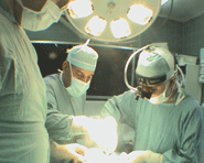
Bifrontal craniotomy
was performed and right subfrontal approach was
performed initially, to see the right optic
nerve and mobilizing during that the right
olfactory tract.
The cystic mass was attacked
lateral to the right optic nerve. Around 70 ml
of yellowish milky fluid was evacuated. The
walls of a craniopharyngioma was removed from
the medial side of the right optic nerve, which
was pushed anteriorly. All the material was sent
for histologic studies.
So as to assure that there is
no remnant of the tumor the other optic nerve
was seen running under the lesion. Part of the
left olfactory tract was mobilized to prevent
traction avulsion. Both ICAs and the basilar
artery were inspected without touching them to
avoid the possible progression of vasospasm.
After dissecting all the necessary neural and
vascular structures with anatomical
preservation, it was sure that total resection
of the craniopharyngioma was achieved.
Routine closure of the wound
and smooth postoperative recovery.
The patient start to talk and
moving all limbs and all neurological deficits
regressed immediately after the operation. She
was sent to the ICU for observation.
Comments
The giant dimension of the
tumor made it impossible to see the origin of
its growth. Mostly it was arising from the
pituitary stalk with minimal solid component.
Her neurologic manifestations were due to
hypertensive-diencephalic syndrome, which
resolved dramatically after the operation.
Bifrontal craniotomy give 160
degrees angle of vision. The lower edge of the
bone defect must be flush with the anterior
fossa floor, even violating the frontal sinuses,
as in this case.
Mobilizing the olfactory
tracts make it possible to preserve them
anatomically and in 85% functionally.
By this method, the surgeon
have almost an absolute visual control about
what he is doing in the sellar region.
For more details about
craniopharyngiomas, please click
here
or
here!
Please! wait for 3-5 min till the
video start to load. It depends upon the internet
connection.