Inomed Stockert Neuro N50. A versatile
RF lesion generator and stimulator for
countless applications and many uses
Multigen RF lesion generator .
26-NOVEMBER-2019 ALI HASAN ALABED ALRAS 50 YEARS
RECURRENT EXTERNAL BLEEDING FROM THE RIGHT CCA AND ECA .
Anamnesis
The patient was operated by me
15-January-2015 for complete occlusion of
the right ICA for repeated CVA attacks with left
sided plegia. The patient came for follow up and
MRA of the carotids performed 03-March-2015
showed acceptable circulation of the right ICA
with improvement of the patient neurologic
status. The patient then came 07-March-2019
telling that he got enlargement under the old
incision site pulsating with pain at the
lesion for the last 10 days. The patient was
neurologically free. He was sent for
investigations. MRA of the carotids showed
pseudo-aneurysm with dissection 16.8x7.4 mm at
the right ICA with clot 41.5x21.4 mm
multilobulated lateral to the artery. The right
extracranial ICA is not seen but the cross
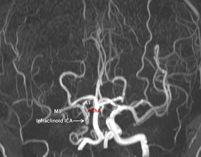
circulation is filling the right infraclinoid,
clinoid, supraclinoid, M1 and A1 from the left
side.
The patient then came 01-May-2019 with oozing of
the mass after manipulation by a doctor. The
patient was advised to keep in conservative
treatment. The patient then came
03-November-2019 telling that yesterday, he
progressed loss of vision right eye for 30
minutes then recovered. He has small clot at the
most upper anterior edge of the previous
incision. He told me that all the period the
wound was quite, and subcutaneous mass appeared
at the center of the incision. It was pulsating,
for what another MRI investigation was performed
and MRA of the right carotid was showing an
aneurysm at the bifurcation of the CCA. The
patient was advised to stop plavix and baby
aspirin and try conservative treatment. The
patient then came to the emergency of Shmaisani
hospital, transferred from other hospital after
resuscitating him from massive bleeding with
hypovolimic shock. When I saw him, he was alert
and the bleeding was stopped and advised to have
blood transfusion with FFP to prepare him to
surgery. Before transfusion the Hb was 10.7
mg/dL.
The old incision was extended
down to expose the right CCA. The vagus nerve was
separated and the CCA was circumscribed by rubber to
protect it in case of urgent escalation of
complications. Using Inomed ISIS Neuroexplorer with
SEP protocol for both hands, the activity of the
brain was monitored during all stages of surgery. A
clamp was applied to the right CCA without
complication. Angiography of the right CCA was
performed and the branches of ECA were seen.
Step-wise upward dissection of the CCA until the
graft was seen There was a huge clot over the upper
and anterior border of the graft. The clot was
removed and the defect of the graft which was
located upper medial was seen. Massive bleeding took
place from the back flow of the right ECA. Using
nylon 4 zero the defect was repaired. The bleeding
stopped and the clamp was removed from the right
CCA. No active bleeding. Routine
closure of the wound.
Smooth postoperative recovery. He was sent to the ward.
The patient is neurologically free walking after 5
hours of surgery.
Comments
This case in one of the most challenging
and difficult to perform.
ISIS SEP is an important part of surgery
to know the condition of the brain after clamping of the
CCA.
Anesthesia protocol is an important part
to make the patient wake during surgery and to see the
movement of the left upper and left lower limbs.
Angiography was important to evaluate the
back flow of the right ECA. In this case, retrospectively,
ligation of the CCA will not resolve his problem, and
massive back flow of the right ECA will trigger second
bleeding attacks.
Exploration of the defect and removing
the clot and repairing the defect with 4 zero nylon, not
only resolved the essential problem, but also made
unnecessary to legate the right CCA.
Skyra MRI with all clinical applications in the run since 28-Novemeber-2013.
Inomed Riechert-Mundinger System, with three point
fixation is the most accurate system in the market. The microdrive and
its sensor gives feed back about the localization.
Inomed MER system
Leica HM500
The World's first and the only Head mounted Microscope.
Freedom combined with Outstanding Vision, but very bad video recording and
documentation.
After long years TRUMPF TruSystem 7500 is running with in the neurosuite at
Shmaisani hospital starting from 23-March-2014
Fig:-1. The right ICA receiving cross circulation from the left
side.
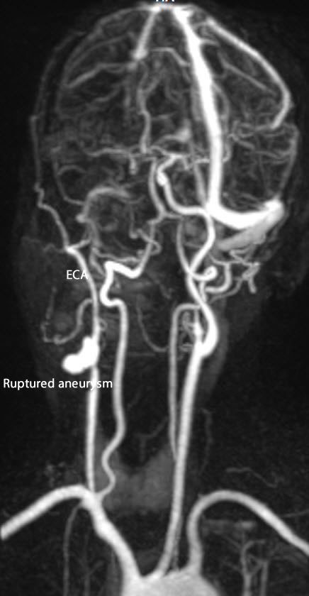
Fig-2: The ruptured aneurysm with external bleeding caused
hypovolimic shock with absent right ICA and preserved ECA.
Notice: Not all operative activities
can be recorded due to lack of time.
Notice: Head injuries and very urgent surgeries are also
escaped from the plan .