Most of the site will reflect the ongoing surgical activity of Prof. Munir Elias MD., PhD. with brief slides and weekly activity.

Functional Neurosurgery
functionalneuro.surgery
Functionalneurosurgery.net

IOM Sites
iomonitoring.org
operativemonitoring.com

Neurosurgical Sites
neurosurgery.art
neurosurgery.me
neurosurgery.mx
skullbase.surgery

Neurosurgical Encyclopedia
neurosurgicalencyclopedia.org

Neurooncological Sites
acousticschwannoma.com
craniopharyngiomas.com
ependymomas.com
gliomas.info
meningiomas.org
neurooncology.me
pinealomas.com
pituitaryadenomas.com 

Neuroanatomical Sites
humanneuroanatomy.com 
microneuroanatomy.com

Neuroanesthesia Sites
neuro-anesthessia.org

Neurobiological Sites
humanneurobiology.com

Neurohistopathological
neurorhistopathology.com

Neuro ICU Site
neuroicu.info

Neuroophthalmological
neuroophthalmology.org

Neurophysiological Sites
humanneurophysiology.com

Neuroradiological Sites
neuroradiology.today

NeuroSience Sites
neuro.science

Neurovascular Sites
vascularneurosurgery.com

Personal Sites
cns.clinic

Spine Surgery Sites
spine.surgery
spondylolisthesis.info
paraplegia.today

Stem Cell Therapy Site
neurostemcell.com


Inomed Stockert Neuro N50. A versatile
RF lesion generator and stimulator for
countless applications and many uses
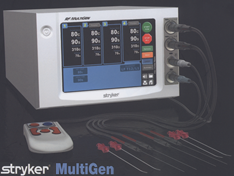

Multigen RF lesion generator .


PERCUTANEOUS TRIGEMINAL NERVE COMPRESSION.


Percutaneous Trigeminal Nerve Compression

Percutaneous trigeminal nerve compression (PTC) has now been convincingly developed, and results of its use will be analyzed in detail because it seem to add another category of patients with trigeminal neuralgia whose treatment by a relatively minor procedure in the middle fossa yields relief. The crucial point is that this relief is accompanied in many by little or no sensory loss immediately after operation, or recovery to this status within a few months.

Rationale for the Procedure

In 1983 Mullan and Lichtor introduced a percutaneous version of Shelden's 1955 open middle fossa exposure for deliberate rubbing or vigorous massage of the trigeminal ganglion and rootlets. Concerning the latter, only the fact that four patients developed anesthesia dolorosa among 1590 reported cases in 28 publications was a vast improvement over the results with trigeminal rhizotomy. Those four cases were by a surgeon (Olivecrona) who compressed with enough vigor to produce some sensory loss in follow­ups at 6 to 8 years in 52 of 94 patients. A facial paralysis, usually temporary, was the most common non trigeminal sequel. The disadvantage of a 25 to 35 percent rate of recurrence of severe pain was not forbidding if mortality and major morbidity could be eliminated by a truly safe percutaneous technique.

Operative procedure

Mullan's thoughtfully developed tactic is to pass a 14-gauge needle to but not through the foramen ovale and to inflate a Fogarty catheter's balloon placed into Meckel's cave to about 10 mm behind the foramen ovale. This has proved to be a rational major improvement on the results of the open operation and to have important advantages over the other two percutaneous procedures. With the patient under general anesthesia the balloon is inflated with 0.5 to 1.0 ml of a water-soluble contrast medium and observed fluoroscopically to make sure the expansion is primarily in the posterior part of the middle cranial fossa, preferably with a small posterior nubbin bulging through the porus trigemini into the posterior fossa. Frequent observation is needed to detect promptly (1) a sudden disruption of the dural sheath of the ganglion with enlargement of the balloon, (2) migration of the balloon into the posterior fossa, or (3) rupture of the balloon. None of these problems caused a lasting sequel. For example, Fraioli's balloons broke six times without causing any complication. However, in Lobato's four cases, when the balloon moved into the posterior fossa for 15 to 30 sec a diplopia lasting 1 week to 4 months ensued. Abdennebi and Amber, having cut two balloons with the sharp edge of a trocar or needle, recommend enveloping the metal with a Teflon catheter. Lichtor and Mullan found that 1 min of inflation sufficed to control the pain and reduced the incidence of persistent dysesthesia. To see one of the procedures click here!

Pressure in the Balloon: Degree and Duration

There is a little uncertainty as to the proper degree and duration of balloon pressure. Lobato et al. infrequently exceeded 1 min. As noted, Lichtor and Mullan decided after 60 cases to drop the duration to 1 min. Likewise, Fraioli et al. discontinued their 3- to 7-min range in favour of 1 to 2 min of inflation. The reverse tactic has been adopted by the Abdennebi and Meglio groups. The former tried initially a range from 4 to 9 min for 20 cases, adopting the 7- to 9-min range for the last 30. The most striking differences between short and longer compressions were recorded by Meglio et al. with only 76 percent of 21 patients securing immediate pain relief and 84 percent experiencing recurrence after balloon compression of 1 to 3 min. All 53 of their patients undergoing compression for 4 to 10 min had immediate relief and in only 57 percent had the pain recurred. Their high recurrence rates are in some measure explained by the inclusion therein of minor medically controllable pains.

The logical approach of measuring the intraballoon pressure has been taken by three groups, especially when it became apparent that there are substantial variations in the force required to distend the balloons. Zanusso's 22 patients were classified in three groups. The three patients with pressures of 0.9 to 1.3 bars had only temporary relief of pain and no sequelae; the 11 at 1.4 to 1.8 bars were all initially relieved, but 1 had a recurrence and 4 had sequelae; the 8 at 1.9 to 2.4 bars all remained relieved, but all had sequelae (1 bar = 760 mmHg). Lobato's group measured this pressure in 22 of their 144 cases, correlating the appearance of the lateral radiographs therewith. Intraluminal pressures of 700, 1150, and 1700 mmHg were seen with, respectively, (a) no bulge toward the posterior fossa, (b) a bulge with a pear shape, i.e., a posterior nipple, and (c) a marked bulge into posterior fossa producing "intense hemifacial numbness." Brown and Preul found that the recommended pear shape developed at a mean of 815 mmHg, but this ranged from 459 to 1273 mmHg. Lobato et al. say that pressures under 600 mmHg are going to fail. Clearly more data are needed to determine whether or not these measurements can teach us to improve the precision of the operation. Lobato relied on the degree to which the balloon adopts a "fully developed" pear shape, stating that this configuration "almost invariably resulted in long-lasting control of neuralgia." Mullan and Lichtor, placing emphasis on avoidance of dysesthesia, prefer to distend only "until the balloon begins to assume a pear shape," . indicating that it is beginning to protrude out of the cave toward the posterior fossa.

Results of Others

Emphasizing their endorsement of this procedure. three other groups, led by Fraioli, Lobato. and Meglio. have each published paper pointing out continued satisfaction with longer follow-ups. In particular. the incidence of facial dysesthesia severe enough to require medical therapy is less than nearly all of us have reported in large series of percutaneous thermal rhizotomies. The brevity and painlessness of the procedure under continuous general anesthesia and the simplicity of the technique are all important advantages.

Multiple Sclerosis

The Lichtor and Mullan cases include five patients with multiple sclerosis. three of them with bilateral disease. Of the seven sides treated by PTC there were failures on only two sides. On one of those sides two PTCs failed. each after light compression because of dense numbness produced by PTC on the first side. Fraioli et al. gave early relief in one of three patients but the pain recurred. Multiple sclerosis is also more difficult to treat by thermal rhizotomy than is "essential" trigeminal neuralgia.

Repetition of PTC

Repetition of PTC after a late recurrence yielded relief in 34 of 54 operations as follows: Abdennebi and Amber. 7 of 16: Esposito et al.. 7 of 18: Fraioli et al.. 8 of 12: Lichtor and Mullan. 4 of 8: Lobato et al.. 8 of 11: and Peragut et al.. 8 of 9.

Major Sequelae

There have been two serious extratrigeminal sequelae. Dan has kindly described a man with persisting pain following severe V1 and V2 facial injuries without cranial abnormality. In the hope of achieving adequate numbness of the painful area the properly placed balloon was inflated for 12 min. This was without incident until 8 h later when complete blindness came on in the ipsilateral eye. Optic atrophy continues at 2 years. The cause of this loss has not been determined by ophthalmic. angiographic. and computed tomography (CT) studies.

The only death following this procedure. described by Spaziente et al.. occurred in a 62-year-old man with typical trigeminal neuralgia and a moderate asymptomatic hydrocephalus. A larger than usual ( 12-gauge) needle "was not inserted beyond the foramen ovale" but clear CSF emerged when the stylet was removed. This stopped when the needle was retracted slightly and 0.7 ml of nonionic contrast medium injected to inflate the balloon to a characteristic pear shape for 6 min. yielding minimal sensory loss and lasting complete relief of pain. However. headache. drowsiness. and vomiting signalled a subarachnoid hemorrhage shown by CT the next day to fill the basal and sylvian cisterns and to increase the size of the ventricles. Cerebral angiography and studies of blood coagulopathy were normal. After a variety of vicissitudes, death finally ensued 7 months later. There seems to have been no diagnostic or technical error in either of these patients. They illustrate the point that it is well advised, when to submit patients to any invasive procedure only when they have explicitly consented to it in writing with the understanding that any conceivable disaster may happen to them.

Arterial Injuries

The 14-gauge needle is a sizable spear to insert through the facial soft tissue. Lichtor and Mullan emphasize the importance of keeping it out of the intracranial cavity. Their assiduous analyses led in one patient with a fourth nerve paralysis of 3 months duration to the demonstration of a very small dural AVM. and in another patient to an extracranial AVM. In a third case a maxillary artery fistula caused a persistent bruit . "easily eliminated by endovascular wire coil occlusion." Revuelta et al. have added another case of arteriovenous fistula from the middle meningeal artery to the internal jugular vein that closed spontaneously 19 days later as demonstrated by another angiogram. They all suspect that in each case the large needle transfixed a small artery and vein. Lobato et al. were unable to place the balloon in Meckel's cave in one patient who shortly developed a low-flow carotid cavernous fistula that resolved spontaneously in 3 months. Mullan states that if intermittent active bleeding occurs during needle insertion it is desirable to compress above the zygoma as well as below the maxilla. Lobato et al. had arterial bleeding through the cannula in four cases. into the external auditory canal in one. and from the nose in two. They also state that the carotid or accessory meningeal artery traverses the foramen ovale and that a tortuous carotid artery may pass directly over this foramen. They point out a number of other possible bony deficiencies in the region that may permit undesirable arterial puncture. They recommend terminating the procedure if arterial bleeding occurs. with which others agree even when using a 20-gauge needle electrode. Meglio and Cionj report that bleeding not demanding cessation of the procedure occurred in three cases. The one of extracranial origin required both aural and nasal plugging: in another the puncture of the intracranial carotid was thought likely. The bleeding was venous in the third case. The outcome in these patients is not described. There is general agreement that a venipuncture need not stop the operation.

Vasodepressor and Vasopressor Reflexes

Brown and Preul are so impressed with the degrees of bradycardia and hypotension seen during any phase of the procedure in most of the patients that they recommend not only continuous monitoring of arterial blood pressure and heart rate and availability of atropine for intravenous use at once. but also use of a responsive noninvasive temporary cardiac pacemaker throughout the procedure. This vasodepressor reflex was converted to a vasopressor response in four patients by blocking the third trigeminal division with lidocaine. Belber and Rak also advocate intravenous atropine. having used thoracic pressure resuscitation once in a patient with a 15-sec apnoea. Lichtor and Mullan describe several such episodes "with­out incident. " Lobato et al.. on the other hand. noted "a steep increase in blood pressure in almost every patient upon inflation of the balloon. They counteract this with a predistension bolus of vasodilating sodium nitroprusside.

Other Minor Complications

Despite numerous instances of temporary unilateral masticator weakness, none has been permanent. However, Belber and Rak report one patient in whom a bilateral procedure performed in one sitting was followed by severe masticatory weakness of 8 days duration before the onset of recovery. Frank and Fabrizi are the only ones to state that compression adequate to stop third-division pain is likely to cause excessive damage to the first and second divisions. A few extraocular palsies have all been temporary.

The dangers of manipulation in the mid-face of a 14-gauge needle and of inflating a balloon on the floor of the middle cranial fossa have been proven to be small and the chances of obtaining relief at the price of acceptable abnormal facial sensations are huge. The Mullan procedure is clearly superior to intracisternal glycerol injections; his own results are especially impressive. The advantage of brief general anesthesia and minimal numbers with dysesthesia must be weighed against one fatality from hemorrhage and one case of unilateral blindness in patients treated without technical error.

Procedure when PTC Fails

For those who experienced failure of percutaneous compressions who now wish the operation with the best chance of success, Mullan recommends partial root section of the portio major in the posterior fossa. A survey of the results beginning in 1929 with Waiter Dandy, who introduced that operation, finds them to be inferior to those of radiofrequency thermal rhizotomy, with respect to both relief of the paroxysmal pain and incidence of major extratrigeminal sequelae. Dandy himself gave up his partial division of the portio major because of his recurrences. Recently Klun described a nearly 50 percent recurrence rate following division of one-third of the portio major, the same procedure recommended by Brown and Mullan.

In Sweet's series of over 1000 trigeminal radiofrequency percutane­ous rhizotomies and in at least six other series of about 1000 or more, there have been no deaths. He had seen no permanent disability and no lasting deficit of any neural function other than that of the trigeminal nerve. He had not declined to operate on any patient, even the patient in renal or congestive heart failure. These seven groups have been able to provide pain relief in all but an average of 1 percent of their patients.  No operation in the posterior fossa  can equal this record.

To revert to the principal objective of the relation of the treatment to the cause of trigeminal neuralgia, this operation usually produces a temporary mild decrease or no change in objective tests of facial sensation following a 1-cc balloon inflation for only 1 min of neural compression, yet yields pain control superior or comparable to open vascular decompression in the juxtapontine region. No evidence that neural indentation in that region is modified by such a moderate manipulation. Why these modest manoeuvres stop the paroxysms of trigeminal neuralgia remains a complete puzzle. However, the fact that this ultra-low-risk manoeuvre has an excellent chance of stopping the pain to eliminate any justification for an open operation in the middle or posterior cranial fossa as the first invasive procedure in the treatment of trigeminal neuralgia.

Conclusions

The safest invasive manipulation is a percutaneous lesion made by (1) radiofrequency heat to produce hypalgesia. or (2) pure glycerol limited to a 10-min exposure if this produces anesthesia, or (3) inflation of a balloon in Meckel's cave for 1 min.

Inasmuch as an unusually conservative approach has an astonishing likelihood of success, it is not crucial at this point to resolve the question as to the cause or causes of the disorder. With respect to the selection of the first invasive treatment, it is immaterial whether the cause is abnormal myelin and/or axons of sensory trigeminal fibers, or significant extrinsic pressure against them, or both, or neither. If the initial conservative effort fails, there are several reasonable, more aggressive tactics to pursue. Clear-cut vascular compression of the trigeminal rootlets in the posterior fossa is a sufficiently uncommon cause of trigeminal neuralgia that an operation in the posterior fossa is not justified as the first invasive procedure in the treatment of this disorder.

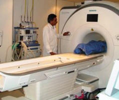
Skyra MRI with all clinical applications in the run since 28-Novemeber-2013.
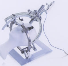

Inomed Riechert-Mundinger System, with three point fixation is the most accurate system in the market. The microdrive and its sensor gives feed back about the localization.
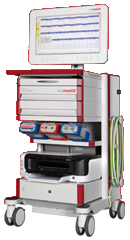

Inomed MER system
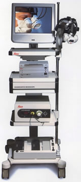
Leica HM500

Leica HM500
The World's first and the only Headmounted Microscope.
Freedom combined with Outstanding Vision, but very bad video recording and documentation.
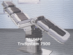
TRUMPF TruSystem 7500

After long years TRUMPF TruSystem 7500 is running with in the neurosuite at Shmaisani hospital starting from 23-March-2014


Back Up!

Notice: Not all operative activities can be recorded due to lack of time.
Notice: Head injuries and very urgent surgeries are also escaped from the plan .


WELCOME TO AL-SHMAISANI HOSPITAL


View Larger Map
 

© [2016] [CNS CLINIC - NEUROSURGERY - JORDAN]. All rights reserved